Anatomical structure of the ear. Bone auditory canal
The ear is a complex organ in humans and animals, through which sound vibrations are perceived and transmitted to the main nerve center of the brain. The ear also performs the function of maintaining balance.
As everyone knows, the human ear is a paired organ located deep in the temporal bone of the skull. Externally, the ear is limited by the auricle. It is the direct receiver and conductor of all sounds.
The human hearing aid can perceive sound vibrations whose frequency exceeds 16 Hertz. The maximum ear sensitivity threshold is 20,000 Hz.
Structure of the human ear
The human hearing system includes:
- External part
- middle part
- Interior
In order to understand the functions performed by certain components, it is necessary to know the structure of each of them. Quite complex sound transmission mechanisms allow a person to hear sounds in the form in which they come from the outside.


- Inner ear. It is the most complex component of the hearing aid. The anatomy of the inner ear is quite complex, which is why it is often called the membranous labyrinth. It is also located in the temporal bone, or more precisely, in its petrous part.
The inner ear is connected to the middle ear through oval and round windows. The membranous labyrinth includes the vestibule, cochlea, and semicircular canals filled with two types of fluid: endolymph and perilymph. Also in the inner ear is the vestibular system, which is responsible for a person’s balance and his ability to accelerate in space. The vibrations that arise in the oval window are transferred to the liquid. With its help, the receptors located in the cochlea are irritated, which leads to the formation of nerve impulses.
The vestibular apparatus contains receptors that are located on the cristae of the canals. They come in two types: cylinder and flask. The hairs are opposite each other. Stereocilia during displacement cause excitation, and kinocilia, on the contrary, contribute to inhibition.
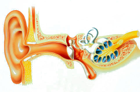
For a more accurate understanding of the topic, we bring to your attention a photo diagram of the structure of the human ear, which shows the complete anatomy of the human ear:

As you can see, the human hearing system is a rather complex system of various formations that perform a number of important, irreplaceable functions. As for the structure of the outer part of the ear, each person may have individual characteristics that do not harm the main function.
Hearing aid care is an integral part of human hygiene, since functional disorders may result in hearing loss, as well as other diseases associated with the outer, middle or inner ear.
According to scientific research, a person experiences vision loss more difficult than hearing loss, because he loses the ability to communicate with the environment, that is, he becomes isolated.
1. Vestibulocochlear organ, organum vestibulocochleare. The structure of the balance organ (pre-cochlear organ).
2. Embryogenesis of the organ of hearing and gravity (balance) in humans.
3. External ear, auris externa. Auricle, auricula. External auditory canal, meatus acusticus externus.
4. Eardrum, membrana tympani. Vessels and nerves of the external ear. Blood supply to the external ear.
5.
6. Auditory ossicles: Hammer, malleus; Anvil, incus; Stirrup, stapes. Functions of the bones.
7. Muscle tensor tympani, m. tensor tympani. Stapedius muscle, m. stapedius Functions of the muscles of the middle ear.
8. Auditory tube, or Eustachian tube, tuba auditiva. Vessels and nerves of the middle ear. Blood supply to the middle ear.
9. Inner ear, labyrinth. Bone labyrinth, labyrinthus osseus. vestibule, vestibulum.
10. Bone semicircular canals, canales semicirculares ossei. Snail, cochlea.
11. Membranous labyrinth, labyrinthus membranaceus.
12. Structure of the auditory analyzer. Spiral organ, organon spirale. Helmholtz's theory.
13. Vessels of the inner ear (labyrinth). Blood supply to the inner ear (labyrinth).
Middle ear, auris media. Tympanic cavity, cavitas tympanica. Walls of the tympanic cavity.
Middle ear, auris media, comprises tympanic cavity And auditory tube connecting the tympanic cavity with the nasopharynx.
Tympanic cavity, cavitas tympanica, lies at the base of the pyramid of the temporal bone between the external auditory canal and the labyrinth (inner ear). It contains a chain of three small bones that transmit sound vibrations from the eardrum to the labyrinth.
Tympanic cavity has a very small size (volume about 1 cm3) and resembles a tambourine placed on its edge, strongly inclined towards the external auditory canal. There are six walls in the tympanic cavity:
1. Lateral wall of the tympanic cavity, paries membranaceus, formed by the eardrum and the bony plate of the external auditory canal. The upper dome-shaped expanded part of the tympanic cavity, recessus membranae tympani superior, contains two auditory ossicles; the head of the malleus and the incus. In case of disease, pathological changes in the middle ear are most pronounced in this recessus.
2. Medial wall of the tympanic cavity adjacent to the labyrinth, and therefore is called labyrinthine, paries labyrinthicus. It has two windows: round, snail window - fenestra cochleae, leading into the cochlea and tightened membrana tympani secundaria, And oval, window of the vestibule - fenestra vestibuli, opening in vestibulum labyrinthi. The base of the third auditory ossicle, the stapes, is inserted into the last hole.
3. Posterior wall of the tympanic cavity, paries mastoideus, carries eminence, eminentia pyramidalis, for premises m. stapedius. Recessus membranae tympani superior continues posteriorly into the mastoid cave, antrum mastoideum, where the airways open cells of the latter, cellulae mastoideae.
Antrum mastoideum is a small cavity protruding towards the mastoid process, from the outer surface of which it is separated by a layer of bone bordering the posterior wall of the auditory canal immediately behind the spina suprameatica, where the cave is usually opened during suppuration in the mastoid process.
4. Anterior wall of the tympanic cavity is called paries caroticus, since the internal carotid artery is close to it. At the top of this wall is internal opening of the auditory tube, ostium tympanicum tubae auditivae, which gapes widely in newborns and young children, which explains the frequent penetration of infection from the nasopharynx into the middle ear cavity and further into the skull.
The outer ear consists of the pinna and the external auditory canal.
Auricle(auricula) has a complex configuration and is divided into two sections: the lobe, which is a duplicate of the skin with fatty tissue inside, and a part consisting of cartilage, covered with thin skin. While it is possible to gather the skin into a fold on the posterior surface, this cannot be done on the anterior surface due to the strong fusion of the skin with the perichondrium. The auricle has a helix (helix), an antihelix (anthelix), a tragus (tragus), an antitragus (antitragus) and an earlobe (lobulus). The tragus covers the entrance to the external auditory canal (Fig. 151).
Rice. 151. Structure of the auricle.
1. Triangular fossa; 2.Rook; 3. Legs of the antihelix; 4. Helix leg; 5.Curl; 6. Shell cavity; 7.Anti-curl; 8. Tragus; 9. Antitragus. 10.Lobe.
Pressure on the tragus area can be painful during an inflammatory process in the external auditory canal, and in children with acute otitis media, since in early childhood the external auditory canal does not have a bony part and is therefore shorter. Pressing on the tragus in these cases leads, in fact, to pressing on the inflamed eardrum, which is accompanied by increased pain. In addition to the indicated protrusions on the front surface of the auricle, there are depressions - a triangular fossa (fossa triangularis), a scapha (scapha). It is necessary to know about these elements of the auricle in order to localize certain processes in the area of the auricle: hematoma in the area of the triangular fossa, abscess of the lobe, etc. It is believed that the height of the auricle normally corresponds to the length of the back of the nose. Deviation in one direction or another allows us to talk about microtia or macrotia. The fact that the auricle is distant from the surface of the skull and has particular blood supply (on the anterior surface of the auricle the vessels are not surrounded by subcutaneous tissue) creates conditions for frostbite, since the vessels are in a state of spasm under the influence of cold. The auricle plays an important role in ototopics, i.e. ability to determine the direction of the sound source, performs a protective function. The normal auricle, due to its complex profile, helps to retain dust particles in the outermost part of the ear canal. When the shell is deformed or completely lost, dust reaches the eardrum and, deposited on it, can contribute to the development of inflammation. The auricle to a certain extent influences the acuity of hearing, therefore, to perceive a weak sound, a person places his palm on the auricle, as if increasing its area.
The auricle, narrowing funnel-shaped, passes into external auditory canal, consisting of two sections: external membranocontilaginous and internal bone(152) . The diameter of the external auditory canal varies, but this does not affect hearing acuity. In children of the first year of life, the bony part of the external auditory canal is absent, and only the cartilaginous part exists. The length of the external auditory canal in children is 0.5-0.7 cm, in adults it is about 3 cm.
Fig. 152. External auditory canal.
The cartilaginous part of the auditory canal, consisting partly of cartilaginous tissue, borders below with the capsule of the parotid salivary gland. The lower wall has several transversely running slits in the cartilaginous tissue. Through them, the inflammatory process can spread to the parotid gland. In the cartilaginous section there are many glands that produce earwax, and there are also hairs with hair follicles, which can become inflamed when pathogenic flora penetrates, resulting in a boil in the external auditory canal.
The cartilaginous section of the external auditory canal is represented by a groove made of cartilage. This groove is open in the area of the posterior-superior wall and therefore, incisions of the ear canal during surgical interventions on the ear, in order to avoid the occurrence of perichondritis, should be made along the posterior-superior wall.
The anterior wall of the external auditory canal closely borders the temporomandibular joint and with each chewing movement this wall moves. In cases where a boil develops on this wall, each chewing movement increases the pain. Close contact of the external auditory canal with the temporomandibular joint causes a fracture of the anterior wall of the auditory canal upon impact in the chin area with skin rupture and possible cicatricial obliteration of the lumen of the auditory canal. In addition, the close anatomical relationship of these formations explains the occurrence of some syndromes related to otorhinolaryngology and dentistry. The bony part of the external auditory canal is lined with thin skin; there is a narrowing at the border with the cartilaginous part. Pushing foreign bodies past this narrowing makes it much more difficult to remove them using one method or another.
The upper wall of the bony part borders on the middle cranial fossa, the posterior wall borders on the air cells of the mastoid process and, in particular, on the cave. This circumstance explains the occurrence of one of the pathognomonic symptoms of an acute inflammatory process in the mastoid process (mastoiditis) - a symptom of overhang of the posterior superior wall in the bony part of the auditory canal, which leads to a narrowing of its lumen due to developing periostitis.
The skin of the external auditory canal in the cartilaginous part is supplied with hair, sebaceous and sulfur glands. The latter secrete sulfur and are modified sebaceous glands. In the bony part of the external auditory canal, the skin is thin and devoid of hair and glands.
Blood supply The external ear is supplied by branches of the external carotid artery. The auricle is supplied with blood from posterior ear And superficial temporal arteries(a. auricularis posterior et a. temporalis superficialis). These same vessels, as well as the deep auricular artery ( a.auricularis profunda) provide blood to the deeper parts and the eardrum, forming a plexus around the external auditory canal.
Venous outflow occurs anteriorly in posterior mandibular vein(v.retromandibularis) and posteriorly into the posterior ear vein (v. auricularis posterior).
Innervation of the external ear(skin of the external auditory canal, auricle) is carried out from the third branch of the trigeminal, vagus and glossopharyngeal nerves. This causes the occurrence of “referred” pain, for example, with inflammation of the periodontal tissue of the eighth lower tooth, you may feel severe pain in the ear on the corresponding side.
The ear is a paired organ located deep in the temporal bone. The structure of the human ear allows it to receive mechanical vibrations in the air, transmit them through internal media, transform them and transmit them to the brain.
The most important functions of the ear include analysis of body position and coordination of movements.
The anatomical structure of the human ear is conventionally divided into three sections:
- external;
- average;
- internal.
Ear shell
It consists of cartilage up to 1 mm thick, above which there are layers of perichondrium and skin. The earlobe is devoid of cartilage and consists of adipose tissue covered with skin. The shell is concave, along the edge there is a roll - a curl.
Inside it there is an antihelix, separated from the helix by an elongated depression - a rook. From the antihelix to the ear canal there is a depression called the auricle cavity. The tragus protrudes in front of the ear canal.
auditory canal
Reflecting from the folds of the concha of the ear, the sound moves into the auditory ear 2.5 cm in length, with a diameter of 0.9 cm. The basis of the ear canal in the initial section is cartilage. It resembles the shape of a gutter, open upward. In the cartilaginous section there are santorium fissures bordering the salivary gland.
The initial cartilaginous part of the ear canal passes into the bone part. The passage is curved in a horizontal direction; to examine the ear, the shell is pulled back and up. For children - back and down.
The ear canal is lined with skin containing sebaceous and sulfur glands. Sulfur glands are modified sebaceous glands that produce. It is removed by chewing due to vibrations of the walls of the ear canal.
It ends with the eardrum, blindly closing the auditory canal, bordering:
- with the joint of the lower jaw, when chewing, the movement is transmitted to the cartilaginous part of the passage;
- with cells of the mastoid process, facial nerve;
- with the salivary gland.
 The membrane between the outer ear and the middle ear is an oval translucent fibrous plate, measuring 10 mm in length, 8-9 mm in width, 0.1 mm in thickness. The membrane area is about 60 mm 2.
The plane of the membrane is located obliquely to the axis of the ear canal at an angle, drawn funnel-shaped into the cavity. The maximum tension of the membrane is in the center. Behind the eardrum is the middle ear cavity.
There are:
- middle ear cavity (tympanum);
- auditory tube (Eustachian tube);
- auditory ossicles.
Tympanic cavity
The cavity is located in the temporal bone, its volume is 1 cm 3. It houses the auditory ossicles, articulated with the eardrum.
The mastoid process, consisting of air cells, is located above the cavity. It houses a cave - an air cell that serves in the anatomy of the human ear as the most characteristic landmark when performing any operations on the ear.
Eustachian tube
 The formation is 3.5 cm long, with a lumen diameter of up to 2 mm. Its upper mouth is located in the tympanic cavity, the lower pharyngeal mouth opens in the nasopharynx at the level of the hard palate.
The auditory tube consists of two sections, separated by its narrowest point - the isthmus. A bony part extends from the tympanic cavity, and below the isthmus there is a membranous-cartilaginous part.
The walls of the tube in the cartilaginous section are normally closed, opening slightly during chewing, swallowing, and yawning. The expansion of the lumen of the tube is provided by two muscles associated with the velum palatine. The mucous membrane is lined with epithelium, the cilia of which move towards the pharyngeal mouth, providing the drainage function of the pipe.
 The smallest bones in human anatomy, the auditory ossicles of the ear, are intended to conduct sound vibrations. In the middle ear there is a chain: malleus, stirrup, incus.
The malleus is attached to the tympanic membrane, its head articulates with the incus. The incus process is connected to the stapes, which is attached at its base to the window of the vestibule, located on the labyrinthine wall between the middle and inner ear.

The structure is a labyrinth consisting of a bone capsule and a membranous formation that follows the shape of the capsule.
In the bone labyrinth there are:
- vestibule;
- snail;
- 3 semicircular canals.
Snail

The bone formation is a three-dimensional spiral of 2.5 turns around the bone rod. The width of the base of the cochlear cone is 9 mm, the height is 5 mm, the length of the bone spiral is 32 mm. A spiral plate extends from the bone rod into the labyrinth, which divides the bone labyrinth into two channels.
At the base of the spiral lamina are the auditory neurons of the spiral ganglion. The bony labyrinth contains perilymph and a membranous labyrinth filled with endolymph. The membranous labyrinth is suspended in the bony labyrinth using cords.
Perilymph and endolymph are functionally connected.
- Perilymph – its ionic composition is close to blood plasma;
- endolymph - similar to intracellular fluid.
 Violation of this balance leads to increased pressure in the labyrinth.
The cochlea is an organ in which physical vibrations of the perilymph fluid are converted into electrical impulses from the nerve endings of the cranial centers, which are transmitted to the auditory nerve and the brain. At the top of the cochlea there is an auditory analyzer - the organ of Corti.
vestibule
The most ancient anatomically middle part of the inner ear is the cavity bordering the scala cochlea through a spherical sac and semicircular canals. On the wall of the vestibule leading into the tympanic cavity, there are two windows - an oval window, covered by the stapes, and a round window, which represents the secondary eardrum.
Features of the structure of the semicircular canals
All three mutually perpendicular bony semicircular canals have a similar structure: they consist of an expanded and simple pedicle. Inside the bones there are membranous canals that repeat their shape. The semicircular canals and vestibular sacs make up the vestibular apparatus and are responsible for balance, coordination, and determining the position of the body in space.
In a newborn, the organ is not formed and differs from an adult in a number of structural features.
Auricle
- The shell is soft;
- the lobe and curl are weakly expressed and are formed by the age of 4 years.
auditory canal
- The bone part is not developed;
- the walls of the passage are located almost closely;
- The drum membrane lies almost horizontally.
- Almost the same size as an adult;
- In children, the eardrum is thicker than in adults;
- covered with mucous membrane.
Tympanic cavity
 In the upper part of the cavity there is an open gap, through which, in acute otitis media, the infection can penetrate into the brain, causing the phenomenon of meningism. In an adult, this gap closes.
The mastoid process in children is not developed; it is a cavity (atrium). The development of the appendage begins at the age of 2 years and ends by 6 years.
Eustachian tube
In children, the auditory tube is wider, shorter than in adults, and located horizontally.
The complex paired organ receives sound vibrations of 16 Hz - 20,000 Hz. Injuries and infectious diseases reduce the sensitivity threshold and lead to gradual hearing loss. Advances in medicine in the treatment of ear diseases and hearing aids make it possible to restore hearing in the most difficult cases of hearing loss.
Video about the structure of the auditory analyzer