Centers of the parasympathetic system. Sympathetic and parasympathetic divisions of the nervous system
Table of contents of the topic "Autonomic (autonomic) nervous system.":1. Autonomic (autonomic) nervous system. Functions of the autonomic nervous system.
2. Autonomic nerves. Exit points of autonomic nerves.
3. Reflex arc of the autonomic nervous system.
4. Development of the autonomic nervous system.
5. Sympathetic nervous system. Central and peripheral divisions of the sympathetic nervous system.
6. Sympathetic trunk. Cervical and thoracic sections of the sympathetic trunk.
7. Lumbar and sacral (pelvic) sections of the sympathetic trunk.
9. Peripheral division of the parasympathetic nervous system.
10. Innervation of the eye. Innervation of the eyeball.
11. Innervation of the glands. Innervation of the lacrimal and salivary glands.
12. Innervation of the heart. Innervation of the heart muscle. Innervation of the myocardium.
13. Innervation of the lungs. Innervation of the bronchi.
14. Innervation of the gastrointestinal tract (intestine to the sigmoid colon). Innervation of the pancreas. Innervation of the liver.
15. Innervation of the sigmoid colon. Innervation of the rectum. Innervation of the bladder.
16. Innervation of blood vessels. Innervation of blood vessels.
17. Unity of the autonomic and central nervous systems. Zones Zakharyin - Geda.
Parasympathetic part historically develops as a suprasegmental department, and therefore its centers are located not only in the spinal cord, but also in the brain.
Parasympathetic centers
Central part of the parasympathetic division consists of the head, or cranial, section and the spinal, or sacral, section.
Some authors believe that parasympathetic centers are located in the spinal cord not only in the region of the sacral segments, but also in other parts of it, in particular in the lumbar-thoracic region between the anterior and posterior horn, in the so-called intermediary zone. The centers give rise to efferent fibers of the anterior roots, causing vasodilation, delayed sweating and inhibition of contraction of involuntary hair muscles in the area of the trunk and limbs.
Cranial section in turn, consists of centers located in the midbrain (mesencephalic part), and in the rhomboid brain - in the pons and medulla oblongata (bulbar part).
1. Mesencephalic part presented nucleus accessorius n. oculomotorii and the median unpaired nucleus, due to which the muscles of the eye are innervated - m. sphincter pupillae and m. ciliaris.
2. Boulevard part represented by n ucleus saliva tonus superior n. facialis(more precisely, n. intermedius), nucleus salivatorius inferior n. glossopharyngei And nucleus dorsalis n. vagi(see corresponding nerves).
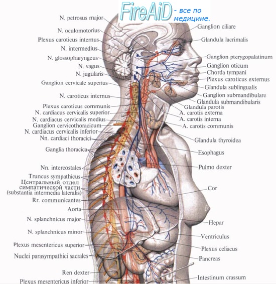
The parasympathetic nervous system consists of central and peripheral sections (Fig. 11).
The parasympathetic part of the oculomotor nerve (III pair) is represented by the accessory nucleus, nucl. accessorius, and the unpaired median nucleus, located at the bottom of the cerebral aqueduct. Preganglionic fibers go as part of the oculomotor nerve (Fig. 12), and then its root, which is separated from the lower branch of the nerve and approaches the ciliary ganglion, ganglion ciliare (Fig. 13), located in the posterior part of the orbit outside the optic nerve. In the ciliary ganglion, the fibers are also interrupted by postganglionic fibers as part of the short ciliary nerves, nn. ciliares breves, penetrate the eyeball to m. sphincter pupillae, ensuring the reaction of the pupil to light, as well as to m. ciliaris, affecting changes in the curvature of the lens.
Fig. 11. Parasympathetic nervous system (according to S.P. Semenov).
SM - midbrain; PM - medulla oblongata; K-2 - K-4 - sacral segments of the spinal cord with parasympathetic nuclei; 1- ciliary ganglion; 2- pterygopalatine ganglion; 3- submandibular ganglion; 4- ear ganglion; 5- intramural ganglia; 6- pelvic nerve; 7- pelvic plexus ganglia; III-oculomotor nerve; VII - facial nerve; IX - glossopharyngeal nerve; X - vagus nerve.
The central division includes nuclei located in the brain stem, namely in the midbrain (mesencephalic region), the pons and medulla oblongata (bulbar region), as well as in the spinal cord (sacral region).
The peripheral department is represented by:
1) preganglionic parasympathetic fibers passing through the III, VII, IX, X pairs of cranial nerves and anterior roots, and then the anterior branches of the II - IV sacral spinal nerves;
2) nodes of the third order, ganglia terminalia;
3) postganglionic fibers, which end on smooth muscle and glandular cells.
Postganglionic sympathetic fibers from the plexus ophthalmicus to m. pass through the ciliary ganglion without interruption. dilatator pupillae and sensory fibers - processes of the trigeminal ganglion, passing through n. nasociliaris for innervation of the eyeball. 
Fig. 12. Scheme of parasympathetic innervation m. sphincter pupillae and parotid salivary gland (from A.G. Knorre and I.D. Lev).
1- endings of postganglionic nerve fibers in m. sphincter pupillae; 2- ganglion ciliare; 3-n. oculomotorius; 4- parasympathetic accessory nucleus of the oculomotor nerve; 5- endings of postganglionic nerve fibers in the parotid salivary gland; 6-nucleus salivatorius inferior;7-n.glossopharynge-us; 8 - n. tympanicus; 9-n. auriculotemporalis; 10-n. petrosus minor; 11- ganglion oticum; 12-n. mandibularis.
Rice. 13. Diagram of connections of the ciliary node (from Foss and Herlinger) 
1-n. oculomotorius;
2-n. nasociliaris;
3- ramus communicans cum n. nasociliari;
4- a. ophthalmica et plexus ophthalmicus;
5-r. communicans albus;
6- ganglion cervicale superius;
7- ramus sympathicus ad ganglion ciliare;
8- ganglion ciliare;
9-nn. ciliares breves;
10- radix oculomotoria (parasympathica).
The parasympathetic part of the interfacial nerve (VII pair) is represented by the superior salivary nucleus, nucl. salivatorius superior, which is located in the reticular formation of the bridge. The axons of the cells of this nucleus are preganglionic fibers. They pass as part of the intermediate nerve, which joins the facial nerve.
In the facial canal, parasympathetic fibers are separated from the facial nerve in two portions. One portion is separated in the form of a large petrosal nerve, n. petrosus major, the other - the drum string, chorda tympani (Fig. 14). 
Rice. 14. Scheme of parasympathetic innervation of the lacrimal gland, submandibular and sublingual salivary glands (from A.G. Knorre and I.D. Lev).
1 - lacrimal gland; 2 - n. lacrimalis; 3 - n. zygomaticus; 4 - g. pterygopalatinum; 5 - r. nasalis posterior; 6 - nn. palatini; 7 - n. petrosus major; 8, 9 - nucleus salivatorius superior; 10 - n. facialis; 11 - chorda tympani; 12 - n. lingualis; 13 - glandula submandibularis; 14 - glandula sublingualis. 
Rice. 15. Diagram of connections of the pterygopalatine ganglion (from Foss and Herlinger).
1-n. maxillaris;
2-n. petrosus major (radix parasympathica);
3-n. canalis pterygoidei;
4-n. petrosus profundus (radix sympathica);
5-g. pterygopalatinum;
6-nn. palatini;
7-nn. nasales posteriores;
8-nn. pterygopalatini;
9-n. zygomaticus.
The greater petrosal nerve departs at the level of the ganglion, leaves the canal through the cleft of the same name and, located on the anterior surface of the pyramid in the groove of the same name, reaches the apex of the pyramid, where it leaves the cranial cavity through the lacerated foramen. In the area of this opening, it connects with the deep petrosal nerve (sympathetic) and forms the nerve of the pterygoid canal, n. canalis pterygoidei. As part of this nerve, preganglionic parasympathetic fibers reach the pterygopalatine ganglion, ganglion pterygopalatinum, and end on its cells (Fig. 15).
Postganglionic fibers from the ganglion as part of the palatine nerves, nn. palatini, are sent to the oral cavity and innervate the glands of the mucous membrane of the hard and soft palate, as well as as part of the posterior nasal branches, rr. nasales posteriores, innervate the glands of the nasal mucosa. A minority of postganglionic fibers reach the lacrimal gland as part of n. maxillaris, then n. zygomaticus, anastomotic branch and n. lacrimalis (Fig. 14).
Another portion of preganglionic parasympathetic fibers as part of the chorda tympani joins the lingual nerve, n. lingualis, (from the III branch of the trigeminal nerve) and as part of it approaches the submandibular node, ganglion submandibulare, and ends in it. The axons of the node cells (postganglionic fibers) innervate the submandibular and sublingual salivary glands (Fig. 14).
The parasympathetic part of the glossopharyngeal nerve (IX pair) is represented by the inferior salivary nucleus, nucl. salivatorius inferior, located in the reticular formation of the medulla oblongata. Preganglionic fibers exit the cranial cavity through the jugular foramen as part of the glossopharyngeal nerve, and then its branches - the tympanic nerve, n. tympanicus, which penetrates the tympanic cavity through the tympanic tubule and, together with the sympathetic fibers of the internal carotid plexus, forms the tympanic plexus, where some of the parasympathetic fibers are interrupted and postganglionic fibers innervate the glands of the mucous membrane of the tympanic cavity. Another part of the preganglionic fibers in the lesser petrosal nerve, n. petrosus minor, exits through the fissure of the same name and along the fissure of the same name on the anterior surface of the pyramid reaches the sphenoid-petrosal fissure, leaves the cranial cavity and enters the ear ganglion, ganglion oticum (Fig. 16). The auricular node is located at the base of the skull under the foramen ovale. Here the preganglionic fibers are interrupted. Postganglionic fibers consisting of n. mandibularis and then n. auriculotemporalis are directed to the parotid salivary gland (Fig. 12).
The parasympathetic part of the vagus nerve (X pair) is represented by the dorsal nucleus, nucl. dorsalis n. vagi, located in the dorsal part of the medulla oblongata. Preganglionic fibers from this nucleus as part of the vagus nerve (Fig. 17) exit through the jugular foramen and then pass as part of its branches to the parasympathetic nodes (III order), which are located in the trunk and branches of the vagus nerve, in the autonomic plexuses of internal organs (esophageal, pulmonary, cardiac, gastric, intestinal, pancreas, etc.) or at the gates of organs (liver, kidneys, spleen). In the trunk and branches of the vagus nerve there are about 1,700 nerve cells, which are grouped into small nodules. Postganglionic fibers of the parasympathetic nodes innervate the smooth muscles and glands of the internal organs of the neck, chest and abdominal cavity to the sigmoid colon. 
Rice. 16. Ear node connection diagram (from Foss and Herlinger).
1-n. petrosus minor;
2- radix sympathica;
3-r. communicans cum n. auriculotemporal;
4-n. . auriculotemporalis;
5-plexus a. meningae mediae;
6-r. communicans cum n. buccali;
7-g. oticum;
8-n. mandibularis.

Rice. 17. Vagus nerve (from A.M. Grinshtein).
1-nucleus dorsalis;
2-nucleus solitarius;
3-nucleus ambiguus;
4-g. superius;
5-r. meningeus;
6-r. auricularis;
7-g. inferius;
8-r. pharyngeus;
9-n. laryngeus superior;
10-n. laryngeus recurrens;
11-r. trachealis;
12-r. cardiacus cervicalis inferior;
13- plexus pulmonalis;
14- trunci vagales et rami gastrici.
The sacral section of the parasympathetic part of the autonomic nervous system is represented by the intermediate-lateral nuclei, nuclei intermediolaterales, of the II-IV sacral segments of the spinal cord. Their axons (preganglionic fibers) leave the spinal cord as part of the anterior roots, and then the anterior branches of the spinal nerves, forming the sacral plexus. Parasympathetic fibers are separated from the sacral plexus in the form of pelvic splanchnic nerves, nn. splanchnici pelvini, and enter the lower hypogastric plexus. Some preganglionic fibers have an ascending direction and enter the hypogastric nerves, superior hypogastric and inferior mesenteric plexuses. These fibers are interrupted in periorgan or intraorgan nodes. Postganglionic fibers innervate the smooth muscles and glands of the descending colon, sigmoid colon, and internal pelvic organs.
The parasympathetic nervous system “balances” the sympathetic. It ensures the adaptation of the eyes to vision at close range, a decrease in heart rate, activation of the secretion of saliva and other digestive juices, as well as increased intestinal motility. The most striking example of the coordinated activity of the parasympathetic and sympathetic systems is their interaction during sexual intercourse.
The central part of the parasympathetic nervous system consists of the head (cranial) region and the spinal (sacral) region. Preganglionic fibers arise from the brainstem as part of the four cranial nerves (oculomotor, facial, glossopharyngeal and vagus) and from the sacral segments of the spinal cord.
The structure of the parasympathetic nervous system (ganglionic neurons and postganglionic fibers are highlighted in red).A) Cranial parasympathetic system. Preganglionic fibers are distributed as part of four cranial nerves:
1. As part of the oculomotor nerve, which forms a synapse with the ciliary ganglion. Postganglionic fibers are responsible for the innervation of the muscles involved in the accommodation reflex - the sphincter of the pupil and the ciliary muscle.
2. As part of the facial nerve, which forms a synapse with the pterygopalatine ganglion (responsible for the innervation of the lacrimal and nasal glands) and the submandibular ganglion (responsible for the innervation of the submandibular and sublingual salivary glands).
3. As part of the glossopharyngeal nerve, which forms a synapse with the ear ganglion (responsible for innervation).
4. As part of the vagus nerve, which forms synapses with extramural (located near the innervated organ) and intramural (located in the wall of the innervated organ) ganglia of the heart, lungs, lower part of the esophagus, stomach, pancreas, gall bladder, small intestine, as well as the ascending and transverse colon.
 Cranial division of the parasympathetic system. E-V-Edinger-Westphal core; PUD - posterior nucleus of the vagus nerve. The explanation of the remaining abbreviations is presented below the figure above (here we will duplicate them).
 RG-ciliary ganglion; SG-cardiac ganglia; IG-intramural ganglia; MG-myenteric ganglia (ganglia associated with the muscular layer of the intestine);
UG-auricular ganglion; TG-pelvic ganglia; KG-pterygopalatine ganglion; PG-submandibular ganglion.
b) Sacral division of the parasympathetic system. Behind the first lumbar vertebra, the sacral segments of the spinal cord form its terminal part, the conus medullaris. The gray matter of the lateral horns of the sacral segments S2, S3 and S4 of the spinal cord gives rise to preganglionic fibers, which, spreading caudally as part of the anterior roots of the spinal cord, pass into the cauda equina.
After leaving the pelvic sacral foramina, some of the fibers branch off and form the pelvic splanchnic nerves. The fibers of the left and right splanchnic pelvic nerves form synapses either with ganglion cells located in the walls of the colon (distal sections) and rectum, or with the pelvic parasympathetic ganglia, located next to the pelvic sympathetic ones described above.
Postganglionic parasympathetic fibers are responsible for the innervation of the detrusor of the bladder, as well as the medial tunica of the internal pudendal artery and its branches going to the cavernous tissue of the clitoris or penis.
Educational video of the anatomy of the autonomic nervous system (ANS)
Click to enlarge
In this article we will look at what the sympathetic and parasympathetic nervous systems are, how they work, and what are their differences. We have previously covered the topic as well. The autonomic nervous system, as is known, consists of nerve cells and processes, thanks to which the regulation and control of internal organs occurs. The autonomic system is divided into peripheral and central. If the central one is responsible for the work of internal organs, without any division into opposite parts, then the peripheral one is divided into sympathetic and parasympathetic.
The structures of these departments are present in every internal organ of a person and, despite their opposing functions, they work simultaneously. However, at different times, one or another department turns out to be more important. Thanks to them, we can adapt to different climatic conditions and other changes in the external environment. The autonomic system plays a very important role; it regulates mental and physical activity, and also maintains homeostasis (constancy of the internal environment). If you rest, the autonomic system engages the parasympathetic system and the number of heartbeats decreases. If you start running and experience heavy physical activity, the sympathetic department turns on, thereby speeding up the heart and blood circulation in the body.
And this is only a small slice of the activity that the visceral nervous system carries out. It also regulates hair growth, contraction and dilation of pupils, the functioning of one or another organ, is responsible for the psychological balance of the individual, and much more. All this happens without our conscious participation, which is why at first glance it seems difficult to treat.
Sympathetic nervous system
Among people who are unfamiliar with the work of the nervous system, there is an opinion that it is one and indivisible. However, in reality everything is different. Thus, the sympathetic department, which in turn belongs to the peripheral, and the peripheral belongs to the autonomic part of the nervous system, supplies the body with the necessary nutrients. Thanks to its work, oxidative processes proceed quite quickly, if necessary, the work of the heart accelerates, the body receives the proper level of oxygen, and breathing improves.

Click to enlarge
Interestingly, the sympathetic division is also divided into peripheral and central. If the central part is an integral part of the work of the spinal cord, then the peripheral part of the sympathetic has many branches and nerve nodes that connect. The spinal center is located in the lateral horns of the lumbar and thoracic segment. The fibers, in turn, extend from the spinal cord (1st and 2nd thoracic vertebrae) and 2,3,4 lumbar vertebrae. This is a very brief description of where the sympathetic system is located. Most often, the SNS is activated when a person finds himself in a stressful situation.
Peripheral department
It is not so difficult to imagine the peripheral part. It consists of two identical trunks, which are located on both sides along the entire spine. They start from the base of the skull and end at the tailbone, where they converge into a single unit. Thanks to the internodal branches, the two trunks are connected. As a result, the peripheral portion of the sympathetic system passes through the cervical, thoracic and lumbar regions, which we will consider in more detail.
- Cervical region. As you know, it starts from the base of the skull and ends at the transition to the thoracic (cervical 1st ribs). There are three sympathetic nodes here, which are divided into lower, middle and upper. All of them pass behind the human carotid artery. The upper node is located at the level of the second and third cervical vertebrae, has a length of 20 mm, a width of 4 - 6 millimeters. The middle one is much more difficult to find, as it is located at the intersections of the carotid artery and the thyroid gland. The lower node has the largest size, sometimes even merging with the second thoracic node.
- Thoracic department. It consists of up to 12 nodes and has many connecting branches. They reach out to the aorta, intercostal nerves, heart, lungs, thoracic duct, esophagus and other organs. Thanks to the thoracic region, a person can sometimes feel the organs.
- The lumbar region most often consists of three nodes, and in some cases has 4. It also has many connecting branches. The pelvic region connects the two trunks and other branches together.
Parasympathetic Division

Click to enlarge
This part of the nervous system begins to work when a person tries to relax or is at rest. Thanks to the parasympathetic system, blood pressure decreases, blood vessels relax, pupils constrict, heart rate slows down, and sphincters relax. The center of this department is located in the spinal cord and brain. Thanks to efferent fibers, the hair muscles relax, sweat secretion is delayed, and blood vessels dilate. It is worth noting that the structure of the parasympathetic includes the intramural nervous system, which has several plexuses and is located in the digestive tract.
The parasympathetic department helps to recover from heavy loads and performs the following processes:
- Reduces blood pressure;
- Restores breathing;
- Dilates blood vessels in the brain and genital organs;
- Constricts the pupils;
- Restores optimal glucose levels;
- Activates the digestive secretion glands;
- Tones the smooth muscles of internal organs;
- Thanks to this department, cleansing occurs: vomiting, coughing, sneezing and other processes.

In order for the body to feel comfortable and adapt to different climatic conditions, the sympathetic and parasympathetic parts of the autonomic nervous system are activated at different times. In principle, they work constantly, however, as mentioned above, one of the departments always prevails over the other. Once in the heat, the body tries to cool itself and actively secretes sweat; when it urgently needs to warm up, sweating is accordingly blocked. If the autonomic system works correctly, a person does not experience certain difficulties and does not even know about their existence, except for professional necessity or curiosity.
Since the topic of the site is dedicated to vegetative-vascular dystonia, you should know that due to psychological disorders, the autonomic system experiences disruptions. For example, when a person has suffered a psychological trauma and experiences a panic attack in a closed room, his sympathetic or parasympathetic department is activated. This is a normal reaction of the body to an external threat. As a result, a person feels nausea, dizziness and other symptoms, depending on. The main thing is that the patient should understand that this is only a psychological disorder, and not physiological deviations, which are only a consequence. This is why medication treatment is not an effective remedy; they only help relieve symptoms. For a full recovery, you need the help of a psychotherapist.
If at a certain point in time the sympathetic department is activated, blood pressure increases, the pupils dilate, constipation begins, and anxiety increases. When the parasympathetic action occurs, the pupils constrict, fainting may occur, blood pressure decreases, excess weight accumulates, and indecision appears. The most difficult thing is for a patient suffering from a disorder of the autonomic nervous system when he has it, since at this moment disorders of the parasympathetic and sympathetic parts of the nervous system are simultaneously observed.
As a result, if you suffer from a disorder of the autonomic nervous system, the first thing you should do is undergo numerous tests to rule out physiological pathologies. If nothing is revealed, it is safe to say that you need the help of a psychologist who will quickly relieve you of your illness.
The autonomic nervous system regulates the activity of organs involved in the implementation of plant functions of the body. It coordinates the work of all internal organs, regulates metabolic and trophic processes and maintains the constancy of the internal environment of the body. The autonomic nervous system innervates the smooth muscles of internal organs and glandular epithelium. It strengthens or weakens the function of organs, as a result of which it changes the tone of the organ. Functionally, the autonomic nervous system consists of two parts: sympathetic and parasympathetic, which function oppositely.
this system consists of a central and peripheral section. The central division of the autonomic nervous system consists of four parts located in different parts of the brain and spinal cord;
1. Mesencephalic part - in the midbrain, parasympathetic nucleus of the oculomotor nerve.
2. Bulbar part - parasympathetic nuclei of VII, IX and X pairs of cranial nerves.
3. Thoracolumbar part - autonomic nuclei located in the lateral intermediate column of the spinal cord at the level of the VIII cervical, all thoracic and two upper lumbar segments.
4. Sacral part - intermedial nuclei located at the level of II - IV sacral segments of the spinal cord. Of these centers, the mesencephalic, bulbar and sacral belong to the parasympathetic, and the thoracolumbar to the sympathetic nervous system. All these centers, in turn, are under the influence of higher vegetative centers located in the hindbrain, cerebellum, diencephalon and telencephalon.
The peripheral part of the autonomic nervous system includes:
1. Autonomic nerves, branches and nerve fibers. Autonomic fibers are divided into prenodal (preganglionic) and postnodal (postganglionic). Prenodal fibers go from the center to the nodes, and postnodal fibers from the node to the organs.
2. Autonomic nerve nodes are divided by location into: prevertebral, paravertebral nodes related to the sympathetic nervous system, as well as intramural and terminal nodes related to the parasympathetic nervous system.
3. Autonomic plexuses, located around the organs and vessels of the thoracic and abdominal cavities.
Difference between the autonomic nervous system and the somatic
1. Somatic nerves exit the brainstem and spinal cord segmentally and maintain segmental distribution. Autonomic nerves emerge from several areas of the brain and spinal cord.
2. In the reflex arc, the processes of motor neurons of the somatic nervous system, leaving the brain, without interruption, go to the muscles. Motor neurons of the autonomic nervous system lie on the periphery in the autonomic ganglia.
3. Somatic nerve fibers are covered with a myelin sheath, while autonomic nerve fibers are covered very thinly or not at all.
4. Somatic nerves innervate striated muscles and sensory organs. Autonomic nerves innervate the smooth muscles of internal organs and blood vessels, as well as glands.
1. The centers of the parasympathetic nervous system are small and scattered. The centers of the sympathetic nervous system are one and occupy a wide area.
2. The sympathetic nervous system innervates all organs and smooth muscles of the eyeball, while the parasympathetic nervous system is absent in the ureter and some large vessels.
3. The sympathetic nerve ganglia are located in front or on the side of the spinal trunk, and the parasympathetic ganglia are located inside the wall of the internal organs or near the organs.
4. The prenodal fibers of the parasympathetic nerves are long, and the postnodal fibers are short. The prenodal fibers of the sympathetic nervous system are short and the postnodal fibers are long.
Central department
This part of the autonomic nervous system represents various structures of the brain. It turns out that it is scattered throughout the entire brain. In the central section, segmental and suprasegmental structures are distinguished. All formations belonging to the suprasegmental department are united under the name hypothalamic-limbic-reticular complex.
Hypothalamus- this is the structure of the brain located in its lower part, at the base. This cannot be said to be an area with clear anatomical boundaries. The hypothalamus smoothly passes into the brain tissue of other parts of the brain.
the activity of the mammary glands (lactation), adrenal glands, gonads, uterus, thyroid gland, growth, fat breakdown, and the degree of skin color (pigmentation) is regulated. All this is possible thanks to the close connection of the hypothalamus with the pituitary gland, the main endocrine organ of the human body.
Thus, the hypothalamus is functionally connected with all parts of the nervous and endocrine systems.
Conventionally, two zones are distinguished in the hypothalamus: trophotropic and ergotropic. The activity of the trophotropic zone is aimed at maintaining a constant internal environment. It is associated with a period of rest, supports the processes of synthesis and utilization of metabolic products. It exerts its main influences through the parasympathetic division of the autonomic nervous system. Stimulation of this area of the hypothalamus is accompanied by increased sweating, salivation, slowing of heart rate, decreased blood pressure, vasodilation, and increased intestinal motility.
Limbic system
This structure includes part of the temporal lobe cortex, hippocampus, amygdala, olfactory bulb, olfactory tract, olfactory tubercle, reticular formation, cingulate gyrus, fornix, and papillary bodies. The limbic system is involved in the formation of emotions, memory, thinking, ensures eating and sexual behavior, and regulates the sleep-wake cycle.
Reticular formation
This part of the autonomic nervous system is called the reticular system because, like a network, it interweaves all the structures of the brain. This diffuse location allows it to participate in the regulation of all processes in the body. The reticular formation keeps the cerebral cortex in good shape, in constant readiness. This ensures instant activation of the desired areas of the cerebral cortex. This is especially important for the processes of perception, memory, attention and learning.
Individual structures of the reticular formation are responsible for specific functions in the body. For example, there is a respiratory center, which is located in the medulla oblongata. If it is affected for any reason, then independent breathing becomes impossible. By analogy, there are centers of cardiac activity, swallowing, vomiting, coughing, and so on. The functioning of the reticular formation is also based on the presence of numerous connections between nerve cells.
Segmental structures
This part of the central part of the visceral nervous system has a clear division into sympathetic and parasympathetic structures. Sympathetic structures are located in the thoracolumbar spinal cord, and parasympathetic structures are located in the brain and sacral spinal cord.
Sympathetic department
Sympathetic centers are localized in the lateral horns in the following segments of the spinal cord: C8, all thoracic (12), L1, L2. Neurons in this area are involved in the innervation of smooth muscles of internal organs, internal muscles of the eye (regulation of pupil size), glands (lacrimal, salivary, sweat, bronchial, digestive), blood and lymphatic vessels.
 Parasympathetic Division
Contains the following structures in the brain:
· accessory nucleus of the oculomotor nerve (nucleus of Yakubovich and Perlia): control of pupil size;
· lacrimal nucleus: accordingly, regulates tear secretion;
· superior and inferior salivary nuclei: provide saliva production;
· dorsal nucleus of the vagus nerve: provides parasympathetic influences on internal organs (bronchi, heart, stomach, intestines, liver, pancreas).
The sacral section is represented by neurons of the lateral horns of segments S2-S4: they regulate urination and defecation, blood flow to the vessels of the genital organs.

Peripheral department
This section is represented by nerve cells and fibers located outside the spinal cord and brain. This part of the visceral nervous system accompanies the vessels, weaving around their wall, and is part of the peripheral nerves and plexuses (related to the normal nervous system). The peripheral department also has a clear division into the sympathetic and parasympathetic parts. The peripheral department ensures the transfer of information from the central structures of the visceral nervous system to the innervated organs, that is, it carries out the implementation of what is “planned” in the central autonomic nervous system.
Sympathetic department
Represented by the sympathetic trunk, located on both sides of the spine. The sympathetic trunk is two rows (right and left) of nerve ganglia. The nodes are connected to each other in the form of bridges, moving between parts of one side and the other. That is, the trunk looks like a chain of nerve lumps. At the end of the spine, two sympathetic trunks unite into one unpaired coccygeal ganglion.
Erythropoiesis.
Occurs in the red bone marrow with the obligatory presence of vitamin B 12, iron and folic acid.
The most important factor stimulating the formation of red blood cells in the bone marrow is erythropoietin. They direct the development of progenitor cells, accelerate hemoglobin synthesis, and promote the release of reticulocytes from the bone marrow. Erythropoietins are produced mainly in the juxtaglomerular apparatus of the kidney, where an inactive form is formed, converted into erythropoietin after interaction with blood plasma proteins. Erythropoietins are also produced by vascular endothelium, liver and spleen cells. The main stimulator of erythropoietin synthesis is hypoxia.
Erythropoiesis is regulated by certain biologically active substances. Thus, androgens, ACTH, growth hormone, thyroxine enhance, and estrogens weaken erythropoiesis.
The normal lifespan of red blood cells in the circulation is about 100-120 days. Therefore, daily, to maintain a stable red blood cell mass, erythropoiesis must replace about 0.8% to 1.0% of circulating red blood cells. Aging red blood cells become increasingly fragile and are eventually removed from circulation by clearance by macrophages, particularly in the spleen. The end product of hemoglobin breakdown in macrophages is bilirubin, which is conjugated in the liver and excreted in bile and urine.
It is imperative to maintain a balance between the rate of red cell production and the rate of loss of red cells from circulation. The process of destruction of red blood cells is called hemolysis.
Types of hemolysis:
Osmotic hemolysis occurs in a hypotonic solution, the osmolality of which is less than that of the red blood cell itself. In this case, according to the laws of osmosis, the solvent (water) moves through the erythrocyte membrane, which is highly permeable to it, into the cytoplasm. Red blood cells swell, and with significant swelling they are destroyed; the blood becomes transparent (“varnish” blood).
Mechanical hemolysis occurs during intense physical effects on the blood. A significant portion of red blood cells are destroyed during prolonged blood circulation in the system of artificial blood circulation machines (CAB). No matter how perfect their physical properties are (strength, elasticity, smoothness of the inner surface), the main factor is missing - the electrostatic forces of repulsion of the endothelium of the vascular wall and red blood cells from each other. It is these forces that, under physiological conditions, prevent mechanical friction of red blood cells and their destruction.
Mechanical hemolysis of canned blood can occur due to improper transportation - rough shaking, etc.
In a healthy person, slight mechanical hemolysis is observed during prolonged running on hard surfaces (asphalt, concrete); during work involving prolonged strong shaking of the body of miners when drilling rock, etc.
Biological hemolysis is associated with the entry into the blood of substances formed in other living organisms: with repeated transfusion of blood incompatible with the Rh factor, with the bite of snakes, poisonous insects, with mushroom poisoning.
Chemical hemolysis occurs under the influence of fat-soluble substances that disrupt the phospholipid part of the erythrocyte membrane - narcotic anesthetics (ether, chloroform), nitrites, benzene, nitroglycerin, aniline compounds, saponins.
Thermal hemolysisoccurs when blood is stored improperly - freezing it and then quickly thawing it. Intracellular crystallization of biological water leads to the destruction of the erythrocyte membrane.
Intracellular hemolysis. Aging red blood cells are removed from the circulating blood and destroyed in the spleen, liver and slightly in the bone marrow by cells of the phagocytic mononucleotide system.
Leukopoiesis.
Leukocytes develop from corresponding progenitor cells in the red bone marrow, while lymphocytes undergo further differentiation in the lymphoid organs. In the regulation of leukopoiesis, by analogy with erythropoiesis, special biologically active substances - leukopoietins - participate. They affect the red bone marrow, increasing the rate of growth and formation of leukocytes depending on age, time of day, food intake, physical activity, pregnancy, emotional stress, exposure to various damaging factors (ultraviolet radiation, infections, etc.). Lymphopoiesis can be stimulated by external factors. For example, bacterial infections are typically associated with an increase in the proportion of neutrophils and monocytes, whereas viral infections increase the proportion of lymphocytes.
An increase in the number of leukocytes in the blood is not necessarily associated with their additional formation: they can be released from peculiar depots of the red bone marrow, spleen, and lungs.
Thrombocytopoiesis.
The number of platelets naturally increases during physical exertion, stress, blood loss and other conditions, and an additional release of platelets from the spleen occurs. This is facilitated by the influence of estrogens, corticotropins, adrenaline, and serotonin. The main regulator of thrombocytopoiesis is thrombocytopoietins. Depending on the site of formation and mechanism of action, short-acting and long-acting thrombocytopoietins are distinguished. The first are formed in the spleen, they enhance the detachment of blood platelets from megakaryocytes and accelerate their entry into the blood. Interleukins can be stimulants. The latter are found in blood plasma and stimulate the formation of platelets in the bone marrow.
Regulation of hematopoiesis.
In addition to the mechanisms of humoral regulation described above (with the help of erythropoietins, etc.), there is the possibility of nervous regulation of this process. Clear facts indicating this have not been found, but it is known that the hematopoietic organs are abundantly innervated and contain a large number of interoreceptors. In addition, the possibility of changing the content of blood cells as a conditioned reflex reaction was shown.
26. Cardiovascular system. The importance of blood circulation in the body. Heart its meaning, position, structure. Heart valves and their role. Heart vessels.
1. The cardiovascular system includes two systems: circulatory (circulatory system) and lymphatic (lymph circulation system). The circulatory system combines the heart and blood vessels - tubular organs in which blood circulates throughout the body. The lymphatic system includes capillaries, vessels, trunks and ducts branched in organs and tissues, through which lymph flows towards large venous vessels. Along the path of lymphatic vessels from organs and parts of the body to trunks and ducts there are numerous lymph nodes related to the organs of the immune system.
The study of the cardiovascular system is called angiocardiology. The circulatory system ensures the delivery of nutrients, regulatory, protective substances, oxygen to tissues, the removal of metabolic products, and heat exchange. This is a closed vascular network that penetrates all organs and tissues, and has a pumping device in the center - the heart.
2. The blood vessels of the body are combined into the large and small circles of blood circulation, and the coronary circle of blood circulation is additionally distinguished.
1) The systemic circulation - the bodily one begins from the left ventricle of the heart. It includes the aorta, arteries of various sizes, arterioles, capillaries, venules and veins. The large circle ends with two vena cavae flowing into the right atrium. Through the walls of the body's capillaries, the exchange of substances between blood and tissues occurs. Arterial blood gives oxygen to tissues and, saturated with carbon dioxide, turns into venous blood. Typically, an arterial type vessel (arteriole) approaches the capillary network, and a venule emerges from it. For some organs (kidney, liver) there is a deviation from this rule. Thus, an artery, the afferent vessel, approaches the glomerulus of the renal corpuscle, and an artery, the efferent vessel, also leaves the glomerulus. The capillary network inserted between two vessels of the same type (arteries) is called the arterial miraculous network. The capillary network is built according to the type of miraculous network, located between the afferent (interlobular) and efferent (central) veins in the liver lobule - the venous miraculous network.
2) Pulmonary circulation – pulmonary circulation begins from the right ventricle. It includes the pulmonary trunk, which branches into two pulmonary arteries, smaller arteries, arterioles, capillaries, venules and veins. It ends with four pulmonary veins flowing into the left atrium. In the capillaries of the lungs, venous blood, enriched with oxygen and freed from carbon dioxide, turns into arterial blood.
3) Coronary circle of blood circulation - cardiac includes the vessels of the heart itself for blood supply to the heart muscle. It begins with the left and right coronary arteries, which arise from the initial part of the aorta - the aortic bulb. Flowing through the capillaries, the blood delivers oxygen and nutrients to the heart muscle, receives metabolic products, including carbon dioxide, and turns into venous blood. Almost all the veins of the heart flow into a common venous vessel - the coronary sinus, which opens into the right atrium. With the weight of the heart being only 1/125-1/250 of the body weight, 5-10% of all blood ejected into the aorta enters the coronary arteries.
3. Heart (cor, Greek cardia) is a hollow fibromuscular organ in the shape of a cone, the apex of which faces down, left and forward, and the base faces upward and backward. It is located in the chest cavity behind the sternum as part of the organs of the middle mediastinum on the tendon center of the diaphragm. The upper border of the heart is located at the level of the upper edges of the cartilages of the third pair of ribs, the right border protrudes 2 cm beyond the right edge of the sternum. The left border runs along an arcuate line from the cartilage of the third rib to the projection of the apex of the heart. The apex of the heart is located in the left fifth intercostal space, 1-2 cm medial to the left midclavicular line. On the heart, there are sternocostal (anterior), diaphragmatic (lower) and pulmonary (lateral) surfaces, right and left edges, coronary and two (anterior and posterior) interventricular grooves. The coronary groove separates the atria from the ventricles, and the interventricular grooves separate the ventricles. Vessels and nerves are located in the grooves. The anterior wall of the right and left atria has a cone-shaped extension facing anteriorly - the right and left ears. Both ears cover the front of the aorta and pulmonary trunk and represent additional reserve cavities. The size of the heart is compared with the size of a given person’s fist (length 10-15 cm, transverse size - 9-11 cm, anteroposterior size - 6-8 cm). The thickness of the wall of the right atrium is less than the thickness of the left atrium (2-3 mm), the right ventricle - 4-6 mm, the left - 9-11 mm.
The weight of the heart of an adult is 0.4-0.5% of body weight (250-350 g). The volume of the heart of adults is 250-35O ml. The human heart has 4 chambers (cavities): two atria and two ventricles (right and left). One chamber is separated from the other by partitions. The longitudinal septum of the heart has no openings, i.e. its right half does not communicate with the left. The transverse septum divides the heart into atria and ventricles. It contains atrioventricular openings equipped with leaflet valves. The valve between the left atrium and the ventricle is bicuspid (mitral), and between the right atrium and the ventricle is tricuspid. The valves open towards the ventricles and allow blood to flow only in that direction. The pulmonary trunk and aorta at their origin have semilunar valves, consisting of three semilunar valves and opening in the direction of blood flow in these vessels.
The wall of the heart consists of three layers: the inner - endocardium, the middle, thickest - the myocardium and the outer - the epicardium.
1) The endocardium lines all the cavities of the heart from the inside, tightly fused with the underlying muscle layer. Consists of connective tissue with elastic fibers and smooth muscle cells, as well as endothelium. The endocardium forms the atrioventricular valves, valves of the aorta, pulmonary trunk, and valves of the inferior vena cava.
2) Myocardium (muscle layer) - the contractile apparatus of the heart, formed by striated cardiac muscle tissue. The muscles of the atria are separated from the muscles of the ventricles by the right and left fibrous rings located around the atrioventricular openings. The muscular tunic of the atria consists of two layers: superficial and deep; it is thinner than the muscular tunic of the ventricles, consisting of three layers: inner, middle and outer. The muscle fibers of the atria do not pass into the fibers of the ventricles; the atria and ventricles contract at the same time.
3) Epicardium – part of the fibrous-serous membrane covering the heart (pericardium). The serous pericardium consists of an internal visceral plate (epicardium), directly covering the heart and tightly connected to it, and an external parietal (parietal) plate, lining the fibrous pericardium from the inside and passing into the epicardium at the point where large vessels depart from the heart. Between the two plates of the serous pericardium - The parietal and epicardium have a slit-like space - the pericardial cavity, lined with mesothelium, which contains a small amount (up to 50 ml) of serous fluid. The pericardium insulates the heart from surrounding organs, protects the heart from excessive stretching, and the serous fluid between its plates reduces friction during cardiac contractions.
The automaticity of heart contractions, regulation and coordination of the contractile activity of the heart is carried out by its conduction system. It is built from special muscle fibers that have the ability to conduct irritations from the nerves of the heart to the myocardium of the atria and ventricles.
4. Inside the heart, due to the existence of valves, blood flows only in one direction. The opening and closing of heart valves is associated with changes in pressure in the cavities of the heart. The role of the heart valves is to ensure the movement of blood in the cavities of the heart in one direction. In some diseases: rheumatism, syphilis, atherosclerosis, the heart valves cannot close tightly enough, heart function is disrupted, and defects occur.
Heart.
The human heart is a hollow muscular organ. A solid vertical partition divides the heart into left and right halves. The horizontal septum, together with the vertical septum, divides the heart into four chambers. The upper chambers are the atria, the lower chambers are the ventricles.
The wall of the heart consists of three layers. The inner layer is represented by the endothelial membrane ( endocardium, lines the inner surface of the heart). Middle layer ( myocardium) consists of striated muscle. The outer surface of the heart is covered with a serous membrane ( epicardium), which is the inner layer of the pericardial sac - the pericardium. Pericardium(heart shirt) surrounds the heart like a bag and ensures its free movement.
Heart valves. The left atrium is separated from the left ventricle bicuspid valve . On the border between the right atrium and the right ventricle is tricuspid valve . The aortic valve separates it from the left ventricle, and the pulmonary valve separates it from the right ventricle.
When the atria contract ( systole) blood from them enters the ventricles. When the ventricles contract, blood is forcefully ejected into the aorta and pulmonary trunk. Relaxation ( diastole) of the atria and ventricles helps fill the cavities of the heart with blood.
The meaning of the valve apparatus. During atrial diastole the atrioventricular valves are open, the blood coming from the corresponding vessels fills not only their cavities, but also the ventricles. During atrial systole the ventricles are completely filled with blood. This prevents the return of blood to the vena cava and pulmonary veins. This is due to the fact that the muscles of the atria, which form the mouths of the veins, contract first. As the cavities of the ventricles fill with blood, the leaflets of the atrioventricular valves close tightly and separate the cavity of the atria from the ventricles. As a result of contraction of the papillary muscles of the ventricles at the time of their systole, the tendon threads of the atrioventricular valves are stretched and do not allow them to turn towards the atria. Towards the end of ventricular systole, the pressure in them becomes greater than the pressure in the aorta and pulmonary trunk. This promotes discovery semilunar valves of the aorta and pulmonary trunk , and blood from the ventricles enters the corresponding vessels.
Thus, The opening and closing of the heart valves is associated with changes in pressure in the cavities of the heart. The significance of the valve apparatus is that it provides blood movement in the cavities of the heart in one direction.
Cardiac cycle and its phases.
There are two phases in the activity of the heart: systole(reduction) and diastole(relaxation). Atrial systole is weaker and shorter than ventricular systole. In the human heart it lasts 0.1-0.16 s. Ventricular systole – 0.5-0.56 s. The general pause (simultaneous diastole of the atria and ventricles) of the heart lasts 0.4 s. During this period the heart rests. The entire cardiac cycle lasts 0.8-0.86 s.
Atrial systole ensures the flow of blood into the ventricles. The atria then enter the diastole phase, which continues throughout ventricular systole. During diastole, the atria fill with blood.
Conduction system of the heart.
In the heart, a distinction is made between working muscles, represented by striated muscle, and atypical, or special, tissue in which excitation occurs and is carried out.
In humans, atypical tissue consists of:
sinoatrial node, located on the posterior wall of the right atrium at the confluence of the superior vena cava;
atrioventricular node(atrioventricular node), located in the wall of the right atrium near the septum between the atria and ventricles;
atrioventricular bundle(bundle of His), extending from the atrioventricular node in one trunk. The bundle of His, passing through the septum between the atria and ventricles, is divided into two legs going to the right and left ventricles. The bundle of His ends in the thickness of the muscles with Purkinje fibers.
The sinoatrial node is the leading node in the activity of the heart (pacemaker), impulses arise in it that determine the frequency and rhythm of heart contractions. Normally, the atrioventricular node and the His bundle are only transmitters of excitations from the leading node to the heart muscle. However, the ability to automaticity is inherent in the atrioventricular node and the His bundle, only it is expressed to a lesser extent and manifests itself only in pathology..
The automaticity of the atrioventricular connection manifests itself only in cases when it does not receive impulses from the sinoatrial node
Atypical tissue consists of poorly differentiated muscle fibers. Nerve fibers from the vagus and sympathetic nerves approach the nodes of atypical tissue. Extracardiac regulatory mechanisms
- This is nervous extracardiac regulation. It is carried out by impulses coming from the central nervous system along the fibers of the vagus and sympathetic nerves.
Parasympathetic fibers: the bodies of 1 neurons, the processes of which make up the vagus nerves, are located in the medulla oblongata. They end in the intramural ganglia of the heart. There are 2 neurons here, the processes of which go to the conduction system, the myocardium and coronary vessels.
Sympathetic fibers: 1st neurons in the lateral horns of the 5 upper segments of the thoracic spinal cord. The processes end in the cervical and upper thoracic sympathetic ganglia. These nodes contain 2 neurons, the processes of which go to the heart. Most of it goes to the heart from the stellate ganglion.
Irritation of the vagus nerves going to the heart inhibits the work of the heart until it stops completely in diastole (Weber brothers, 1845). The first case of detection of the inhibitory influence of nerves in the body.
With severe irritation, the heart stops working for a while. During this period, the excitability of the heart is reduced - a negative bathmotropic effect; the conduction of excitation is slowed down – a negative dromotropic effect. Often there is a complete blockade of excitation conduction in the atrioventricular node.
With prolonged stimulation of the vagus nerve, heart contractions are restored - “the heart escapes from the influence of the vagus nerve.”
Microelectrode leads from single muscle fibers of the atria revealed hyperpolarization of the MP with strong irritation of the vagus nerve.
The influence of sympathetic nerves on the heart was studied by the Zion brothers (1867), then by I.P. Pavlov (1887). The following were identified: a positive chronotropic effect - increased heart rate (Zions - nerves “heart accelerators”); positive dromotropic effect – improvement of the conduction of excitation in the heart; positive bathmotropic effect - increased excitability of the heart; positive inotropic effect - increased heart contractions without a noticeable increase in rhythm (“strengthening nerve” according to I.P. Pavlov).
The mediator acetylcholine, formed at the endings of the vagus nerve, is quickly destroyed by acetylcholinesterase and, therefore, has only a local effect. Norepinephrine, released at the endings of the sympathetic nerves, is destroyed much more slowly and lasts longer. After the cessation of irritation of the sympathetic nerve, increased frequency and intensification of heart contractions persist for some time. Along with the main transmitter, substances with a modulating effect can be released into the synaptic cleft.
Nervous extracardiac regulation has a corrective effect on the rhythm and function of the heart. The rhythm itself originates in the 1st order pacemaker, and nervous influences accelerate or slow down the rate of spontaneous depolarization of pacemaker cells, changing the modes of heart function. According to I.P. Pavlov, trophic stimulation of metabolic processes also takes place.
However, the triggering effects of the central nervous system are also known, when signals arriving along the nerves initiate heart contractions. This is observed in experiments with stimulation of the vagus nerve in a mode close to natural, i.e. in “volleys” (“packs”) of impulses, and not in a continuous stream, as is traditionally done. When the vagus nerve is irritated by “volleys” of impulses, the heart contracts in the mode of these “volleys”. Each “volley” corresponds to one heart contraction. By changing the frequency and characteristics of the “volleys,” you can control the heart rhythm over a wide range.
Reproduction of the central rhythm by the heart dramatically changes the electrophysiological parameters of the activity of the sinoatrial node. When the node operates in automatic mode, as well as when the frequency changes under the influence of normal irritation of the vagus nerve, excitation occurs at one point of the node. In the case of reproduction of the central rhythm, many node cells simultaneously participate in the initiation of excitation.
The signals that ensure synchronous reproduction of the central rhythm by the heart differ in their mediator nature from the generally accepted influences of the vagus nerve. Along with acetylcholine, regulatory peptides of various compositions are released. Those. the implementation of each type of vagus nerve effects is ensured by its own “transmitter cocktail”.
A change in the frequency of sending “packs” of impulses from the cardiac center of the medulla oblongata in humans can be demonstrated in the following study. The person is asked to breathe faster than his heart beats. To do this, he monitors the flashing of the photostimulator light and produces one breath for each flash of light. The photostimulator is set at a frequency higher than the initial heart rate. As a result of this, in the medulla oblongata, excitation is irradiated from the neurons of the respiratory center to the neurons of the cardiac center, and in the cardiac efferent neurons of the vagus nerve, “packs” of impulses are formed in a new rhythm, common to the respiratory and cardiac centers.
In experiments on dogs, rapid breathing is caused by overheating. As soon as the rhythm of rapid breathing becomes equal to the heart rate, both rhythms are synchronized and then increase or decrease in a certain range synchronously. When the vagus nerves are cut or blocked, the effect of rhythm synchronization disappears.
Thus, along with the intracardiac one, there is also a central heart rhythm generator. Under natural conditions, it forms adaptive reactions of the heart, imposing on the heart the rhythm of signals arriving along the vagus nerves. The intracardiac generator ensures that the pumping function of the heart is maintained in the event of the central generator being turned off.
The centers of the vagus and sympathetic nerves are the 2nd (after intracardiac) level in the hierarchy of nerve centers that regulate the functioning of the heart. They integrate influences descending from the higher parts of the brain.
A higher level of the hierarchy is the centers of the hypothalamus. With electrical stimulation of the hypothalamus, reactions of the cardiovascular system are observed, which in severity exceed the reactions that occur under natural conditions. With local point stimulation of some areas of the hypothalamus, changes in rhythm, force of contraction of the left ventricle, degree of relaxation of the left ventricle, etc. were observed. Those. The hypothalamus contains structures that can regulate certain functions of the heart. But under natural conditions, these structures do not work in isolation. The hypothalamus is the executive organ. It provides an integrative restructuring of the functions of the cardiovascular system (and other systems) according to signals coming from the limbic system or neocortex.
Reflex regulation of heart activity.
Reflex reactions that occur when various receptors are stimulated can either inhibit or