IV. Projections of the heart valves and places where they can be heard (cardiac auscultation points)
Palpation, percussion, auscultation are methods of objective examination used by doctors around the world in the diagnostic process various diseases. These methods are used in conjunction with biochemical and other types of tests, instrumental research, they use technologies, of which there are a large number. Interestingly, an objective examination plays a decisive role in making a diagnosis.
Auscultation is the most complete and informative method. It is used for diagnostics in surgery, therapy, obstetrics, and pediatrics. Using this method, listening determines the presence of pneumonia, bronchitis, cardiac defects and many other pathologies in children and adults.
Auscultation of the adult heart
Along with being highly informative, this is also the most difficult method of objective examination. It requires perfect pitch, a sense of rhythm and constant practice, as it has a large number of nuances. Diagnostics in medicine using the method of auscultation makes it possible to determine heart disease and pulmonary pathology early stage development.
Listening to the heart is performed in a lying or standing position. Some diseases are characterized by changes heart rate after the load, so sometimes, for accurate diagnosis, the patient is taken out of a state of physical rest. The auscultation method requires compliance with certain rules:
- isolation from ambient noise;
- listening to the heart is carried out during (if possible), as well as separately during inhalation and exhalation;
- it is necessary to use a phonendoscope and stethoscope for auscultation of high and low tones;
- First of all, they determine the presence and characteristics of sounds in various points, and then listen to pathological or physiological noises.
Heart percussion
Used to determine the boundaries of the organ and absolute cardiac dullness. IN Lately this method fades into the background. Some experts have completely abandoned it, since the results of percussion are not very accurate and have large percentage subjectivism. This method has been replaced by radiography and ultrasound, which give a complete picture of the size and position of the organ.
Palpation of the heart
Widely used in diagnostics. Palpation of the heart is carried out in order to more clearly determine the position and strength by pressing a finger to the corresponding area. Some diseases are characterized by a slight trembling of the chest, or “cat purring syndrome.”
Ability to listen and hear
The heart is not listened to in a chaotic manner. There are projections of cardiac valves on chest. There are four of them in total.
- Mitral - IV rib, to the left of the sternum.
- Aortic - III rib, to the right of the sternum.
- - III intercostal space on the left.
- Tricuspid - IV intercostal space on the right.
However, auscultation points are slightly different from direct projections, as the sound in these places is clearer and more understandable.
- At the top of the heart -
- II intercostal space, from the sternum to the right - aortic.
An important sign serious illnesses is a cardiac murmur, which can be constant or appear after a certain load. You must be able to listen very well and hear all deviations from the normal heart rate. It is important to determine not only the noise, but also the nature and location of its formation. It can appear in systole or diastole.
Not only noise, but also working phases can be pathological or physiological. Auscultation of the heart helps in diagnosis. Listening points are similar to those described above. Maybe education III and IV additional tones that appear in different conditions(time period, first-second share of systole or diastole).

Small heart - big responsibility
Pediatric auscultation is a very important part of diagnosis. A child, especially a small one, due to his age, cannot report his problems. The pediatrician must have keen hearing and high degree literacy, since the sounds of a child’s heart change along with his growth. Functional or pathological murmurs may be detected. It is important to carry out comparative characteristics between the first and second tones in strength or emphasis. Any violation indicates a number of pathological processes in a child's body.
Differential diagnosis of heart diseases in children using the auscultation method
| Heart tone | Place of emphasis | Identified pathology (physiology) |
| First | Top of the heart | The left atrioventricular orifice is narrowed |
| Second | Aorta | Arterial hypertension or physiological characteristic puberty |
| Second | Pulmonary artery | Open ductus arteriosus, stenosis, bicuspid valve insufficiency, atrial or interatrial defect interventricular septum, sclerosis of the pulmonary artery, pulmonary fibrosis, myocarditis with stagnation in the pulmonary circulation |
| First and second | At all points | after stress (physical or psycho-emotional) |
In addition to accents, heart tones may weaken or split. Auscultation objectively characterizes this if the doctor knows how to listen.
Pregnancy and auscultation
The heart leaf is laid down and begins to contract already in the third week of pregnancy, and at six it can be heard on an ultrasound. Diagnosis of the mother and fetus is mandatory throughout the entire period and especially during childbirth. The number and content of tones constantly changes in proportion to intrauterine development.
Auscultation of the fetus is the simplest and most effective method determining its viability. To perform this simple operation, an obstetric stethoscope is required (photo below). If necessary, use a phonendoscope.
You can conditionally divide the entire pregnancy period into several periods (according to the rate of fetal heartbeats, as well as the nature of their fullness).
Interestingly, at the 6th week after conception, the child’s heart rate coincides with the mother’s. The difference can be 3 strokes up or down. Then the number of contractions begins to increase. If we take into account that the heart rate increases by 3 beats every day, then it is permissible to determine the age of the fetus histologically.
After two months of pregnancy, the heart itself is divided by partitions into 4 chambers - atria and ventricles. The adult organ also has a similar structure. At the beginning of the 9th week, the embryo's heart beats approximately 175 beats per minute. Further, the frequency decreases and, starting from the second trimester, 140-160 beats become the norm for the fetus. Any deviations from it indicate hypoxia, with tachycardia characterizing the initial degree of oxygen deficiency, and bradycardia characterizing a severe stage requiring immediate intervention.
Fetal palpation
By palpation in the second half of pregnancy, you can determine the position of the fetus and its individual parts in the uterus. In addition, the gestational age is determined by the height of the uterine fundus, as well as by the baby’s head: if it is pressed tightly to the entrance to the pelvis, these are the first harbingers of labor. In obstetrics, the Leopold method is used, which consists of four main techniques.
Auscultation and childbirth
Deafness of heartbeats can be both a manifestation of pathology and an elementary difficulty in listening. This happens when the mother’s abdominal wall is thickened (obesity), the fetus is abnormally positioned (for example, a posterior view of the occipital or breech presentation), polyhydramnios, etc. Muted heartbeat tone is especially common during the labor period. Diagnosis of the fetal body at this time is of paramount importance.
One of the methods of examining a pregnant woman is palpation. It helps determine the location of the fetus and its presentation. But the same result can be achieved when cardiac auscultation is used to diagnose intrauterine development. Listening points are characteristic. If the heartbeat is detected more clearly above the mother's navel, then the fetus has a breech presentation, if lower - a cephalic presentation. The baby may be hyperactive, turning over from side to side throughout the entire pregnancy. Listening to clear tones at the level of the navel indicates a transverse position.
Auscultation in the diagnosis of pulmonary diseases

Auscultation is a method that plays crucial in making a diagnosis pulmonary diseases. There are regular (or vesicular) breathing and various shapes deviations from the norm. Also characteristic feature various diseases are dry or moist rales, which have certain auscultation characteristics. The auscultation points of the lungs are located symmetrically.
Physiologically altered vesicular respiration
If a person has a well or, conversely, poorly developed muscle mass, there is a hypertrophied adipose tissue, the change in breathing can be either in the direction of weakening or strengthening. Listening takes place using a phonendoscope.
Increased vesicular respiration is typical in childhood. Its other name, which can be heard in medical circles, is puerile. There is one characteristic feature - the same breathing in symmetrical areas on the right and left sides.
Diagnosis of bronchitis by auscultation
Auscultation for bronchitis is carried out in the usual way. When listening to the acute stage, the vesicular type is characteristic. This is the body's reaction to inflammation and narrowing of the bronchioles. Against the background of harsh breathing, dry wheezing is detected, and they can be different in tone, and also resemble a buzzing and whistling. This depends on the size of the bronchi and the degree to which they are filled with secretions. They are clearly audible in both phases of breathing.

As bronchitis progresses, mucus production in respiratory tract increases, and auscultation reveals medium-bubble rales.
It is best to listen to the lungs when the patient is standing. It is necessary to compare the sounds of breathing and wheezing at the same points on the right and left organs. There is a certain sequence of auscultation - auscultation points - lungs.
You need to start from the tops and then examine the front surface, then the side and back. With prolonged bronchitis, additional sounds may be added, for example, crepitus, which indicates the transition of inflammation to the lower respiratory tract.
Auscultation of the lungs is carried out in several stages: during normal and deep breathing and after coughing. The auscultation points that are most “suspicious” to the doctor are examined in particular detail.
Diagnosis of chronic bronchitis is also based on auscultation data and laboratory research biological materials. When listening to the lungs, breathing is determined to be of the vesicular type in the presence of a longer exhalation or hard, as in the acute stage. Sometimes Chronical bronchitis provokes the development of more serious illness- In this case, breathing becomes “cotton”. During an exacerbation, wheezing is heard over the entire surface of the lungs.
Percussion of the lungs

Percussion examination can be carried out in three ways: by tapping directly on the examined area, through a plate or with a finger on a finger. Currently, the last one is most applicable. This method does not require the doctor to have additional equipment, and it is quite informative in examining the lungs.
Percussion can be comparative or have a topographical focus. The most popular is the first option, which is used to identify pathological foci. They are seals, so percussion sound above them is more dull than above healthy lung tissue.
There are a large number of shades and tones extracted when examining sounds. Normally, it should be loud, ringing and prolonged. If deafness, dullness of tone, a metallic tint, box or tympanitis appear, this indicates that the patient has inflammatory or other processes in the lungs that require medical intervention.
Auscultation in the diagnosis of diseases of the gastrointestinal tract
Listening is used as a method for diagnosing a number of diseases. gastrointestinal tract. The examination is carried out by a doctor using a stethoscope or placing the ear to the abdominal wall. Using this method, the presence (absence) of peristalsis in the intestines or stomach is determined.

Auscultation is carried out on a comparative principle, that is, to obtain an adequate picture, it is necessary to listen at different points. The examination should be carried out in silence and, if possible, without pressing on the abdomen.
Palpation of the abdomen
In organ examination abdominal cavity The palpation method provides maximum information. It is performed with gentle pressure on the abdomen. You need to start from the left groin area with warm hands so as not to cause discomfort to the patient. This is required to eliminate reflex tension of the abdominal wall.
The examination is carried out using the method of comparative analysis of the right and left halves from bottom to top. Pressure on the epigastric area is final. Using this, pain is determined, in various organs, tension of the abdominal wall, the presence of fluid in the abdominal cavity (fluctuation syndrome).
Percussion of the abdomen
The percussion method also makes it possible to identify the spleens, since they have an absolutely dull sound (femoral). In addition, by comparing gastric and intestinal tympanitis, the doctor can make a diagnosis of obstruction of any of the sections.
Absolute hepatic dullness is normally determined with right side in the IV intercostal space at the level of the midline of the nipple. If, upon examination of this area, a tympanic sound is detected, this indicates perforation of the organs, that is, there is fluid in the cavity.
Percussion of the spleen has no practical significance: its lower edge can be easily palpated.
From this article you will learn about such an ancient method of studying health conditions as cardiac auscultation. History of the method, basic principles of auscultation and diseases that can be identified or at least assumed using this technique.
Article publication date: 05/22/2017
Article updated date: 05/29/2019
Auscultation, or auscultation, is a method for assessing certain functions human body, based on the analysis of sounds that certain body systems emit during their work. Listening to the heart is not the only point of application of the technique. You can listen or auscultate the vessels, lungs, intestines. Great importance has a technique in obstetrics, since through the anterior abdominal wall the mother can listen to the murmur of placental vessels and fetal heart sounds. The auscultatory method is the basis for measurement blood pressure according to the Korotkov method - the same one that we all use when measuring pressure with a tonometer.
The method of listening was used by even the most ancient healers, but for this they put their ear to the chest, back or stomach of the patient. The father of modern auscultation can rightfully be called the French doctor Rene Layenek, who, observing the rules of decency, could not put his ear to the chest of a young girl. That is why he rolled a piece of paper into a tube, applied it to the heart area and discovered that in this way the audibility of heart sounds increased significantly. It was Layenek who invented the prototype of the modern stethoscope - a tube with which doctors perform auscultation. He also gave the initial foundations to such a concept as heart auscultation points - certain places on the chest, in which certain noises and sounds of each of the organ structures are most clearly heard. We will talk about these points and their meaning below.
Basic rules for cardiac auscultation
A simple method such as listening requires strict rules:
- The doctor should use only his own approved stethoscope. This is why cardiologists and therapists sometimes use one single stethoscope all their lives and will not lend it to anyone.
- The stethoscope must be appropriate for the patient’s age - that is why in pediatrics and neonatology there are special children’s stethoscopes or special attachments for a regular one.
- The attachment to the stethoscope should be warm, like the air in the room.
- The study should be carried out in silence.
- The patient should remove clothing up to the waist.
- The patient mainly stands or sits, the doctor is in a position that is comfortable for him.
- The stethoscope head should fit snugly against the skin.
- If hairline is very pronounced on the patient’s skin, the skin in this area should be wetted or lubricated with liquid oil.

Two heart tones
The heart is a complex organ consisting of muscle fibers, connective tissue framework and valve apparatus. Valves separate the atria from the ventricles, and also the chambers of the heart from large or great vessels, outgoing or coming into the cardiac chambers. This entire complex structure is constantly in motion, contracting and relaxing rhythmically. Valves open and close, blood moves in impulses inside the vessels and chambers of the organ. Each element of the heart creates certain sounds, united by doctors into the concept of heart sounds. There are two main heart sounds: the first (systolic) and the second (diastolic).

First tone
The first heart sound occurs at the moment of its contraction - systole - and is formed by the following mechanisms:
- Valve mechanism - slamming and corresponding vibration of the bicuspid (mitral) and tricuspid valves, which separate the atria from the ventricles.
- The muscular mechanism is the contraction of the atria and ventricles and the pushing of blood further along the course of its movement.
- Vascular mechanism - oscillation and vibration of the walls of the aorta and pulmonary artery during the passage of a powerful stream of blood from the left and right ventricles, respectively.
Second tone
This tone occurs at the moment of relaxation of the heart muscle and its rest - diastole. It is not as multicomponent as the first one, and consists of only one mechanism: the valve mechanism - the slamming of the aortic and pulmonary artery valves and their vibration under the pressure of blood.
 Phonocardiogram – recording of vibrations and sounds produced by the activity of the heart and blood vessels
 Technique and points of auscultation of the organ
During listening, the doctor must distinguish and evaluate the following parameters of the heart:
- Heart rate (HR) – normally it varies on average from 60 to 85 beats per minute.
- Rhythm of heart contractions - normally the heart works rhythmically, contracting and relaxing after certain periods of time.
- Sonority or volume of heart sounds - the first and second tones must be of a certain volume. The first tone should be louder than the second, no more than twice. Of course, their sound can be affected not only by diseases, but also by the thickness of the chest, the patient’s weight, and the thickness and massiveness of subcutaneous fatty tissue.
- Integrity of heart sounds - the first and second sounds should be heard completely, without separating or bifurcating.
- The presence or absence of pathological heart tones, noises, clicks, crepitus and other signs of diseases of the heart and other organs.
In order for cardiac auscultation to be correct, it is important to follow a certain sequence of listening to heart sounds. The inventor of the stethoscope, Lainenek himself, developed a certain algorithm for listening to the heart and identified places - listening points - where certain nuances of its work are heard more clearly. Modern diagnostics calls these places the auscultation points of the heart, which we mentioned at the beginning of our article. It is at these points that not only the first and second tones are heard, but each of them is the place of best listening to a certain heart valve
There are five such points in total; they form almost a circle along which the researcher’s stethoscope moves.
- Point 1 is the place at the apex of the heart where the mitral or bicuspid valve separating the left chambers of the heart is most clearly audible. Usually this point is located at the site of attachment to the sternum of the cartilage of the fourth rib on the left.
- The 2nd point is the 2nd intercostal space to the right of the edge of the sternum. In this place, the sounds of the aortic valve, which closes the mouth of the largest artery of the human body, are best heard.
- The 3rd point is the 2nd intercostal space to the left of the edge of the sternum. At this point, the sounds of the pulmonary valve are heard, carrying blood from the right ventricle to the lungs for oxygen enrichment.
- The 4th point is the place at the base of the xiphoid process of the sternum - “under the stomach”. This is the point of best audibility of the tricuspid, or tricuspid, valve of the heart, separating its right halves.
- 5 point, called in medical textbooks Botkin-Erb point - III intercostal space at the left edge of the sternum. This is the site of additional auscultation of the aortic valve.
It is at these points that pathological sounds are best heard, indicating certain disturbances in the functioning of the heart valve apparatus and abnormal blood flows. Experienced doctors also use other points - above large vessels, in the jugular notch of the sternum, axillary region.
What diseases and conditions can be identified using auscultation?
I would like to note that a few decades ago, heart auscultation was one of the few methods for diagnosing diseases of cardio-vascular system. Doctors trusted only their ears and made complex diagnoses, unable to confirm them with any instrumental methods other than an electrocardiogram or chest x-ray.
Modern medicine is equipped with a huge arsenal of methods and technologies, so auscultation has undeservedly faded into the background. In fact it is cheap, accessible and quick way, which allows, in a wide flow of patients, to roughly identify individuals who are subject to a more thorough examination: cardiac ultrasound, angiography, and other modern, but far from cheap, techniques.
So, we list the main characteristics of pathological heart sounds that auscultation of the heart helps to identify.
Change in the sonority of heart sounds
- Weakening of 1 tone is observed with myocarditis - inflammation of the heart muscle, myocardial dystrophy, mitral and tricuspid valve insufficiency.
- Strengthening of the first tone occurs with narrowing mitral valve– stenosis, severe tachycardia and changes in heart rhythm.
- A weakening of the second tone is observed in patients with a drop in blood pressure in the systemic or pulmonary circulation, and malformations of the aorta.
- Strengthening of the second tone occurs with increased blood pressure, thickening of the walls or atherosclerosis of the aorta, stenosis of the pulmonary valve.
- Weakening of both tones is observed in case of obesity of the patient, dystrophy and weak heart function, myocarditis, accumulation of fluid in the cavity of the heart sac after inflammatory process or trauma, severe emphysema.
- An increase in both tones is observed with increased cardiac contractility, tachycardia, anemia, and exhaustion of the patient.

The appearance of heart murmurs
A murmur is an abnormal sound effect superimposed on the heart sounds. The noise always occurs due to abnormal blood flows in the cavities of the heart or as it passes through the valves. Noises are assessed at each of five points, which allows you to determine which valve is not working correctly.
It is important to evaluate the volume, sonority of noises, their prevalence in systole and diastole, duration and other characteristics.

- Systolic murmur, that is, murmur during the first tone, may indicate myocarditis, damage to the papillary muscles, bicuspid and tricuspid valve insufficiency, mitral valve prolapse, stenosis of the aortic and pulmonary valves, ventricular and atrial septal defects, and atherosclerotic changes in the heart.
Systolic murmurs can sometimes be present with MARS or minor anomalies of cardiac development - when there are some abnormalities in the structure of the organ and great vessels. anatomical features. These features do not in any way affect the functioning of the heart and blood circulation, but can be detected by auscultation or ultrasound examinations hearts.
- Diastolic murmur is more dangerous and almost always indicates heart disease. Such noises occur in patients with stenosis of the mitral and tricuspid valves, insufficient function of the aortic and pulmonary valves, tumors.
Abnormal heart rhythms
- The gallop rhythm is one of the most dangerous abnormal rhythms. This phenomenon occurs when heart sounds split and sounds similar to the clatter of hooves “ta-ra-ra”. This rhythm appears in severe cardiac decompensation, acute myocarditis, and myocardial infarction.
- The pendulum rhythm is a two-term rhythm with equal pauses between the 1st and 2nd heart sounds, occurring in patients with arterial hypertension, cardiosclerosis and myocarditis.
- The rhythm of the quail sounds like “sleep in time” and is combined with mitral stenosis, when blood passes through a narrow valve ring with enormous effort.
Auscultation cannot become the main criterion for making a particular diagnosis. It is imperative to take into account the person’s age, the patient’s complaints, characteristics of his body weight, metabolism, and the presence of other diseases. And in addition to listening to the heart, all modern cardiological studies should be applied.
Are you completely unfamiliar with auscultation as an examination method? But you are wrong. You encountered this method already in deep childhood and continue to encounter it to this day. And there is nothing surprising about this. It’s just that the name came to us from eighteenth-century France, when the doctor Rene Laennec in 1816 proposed a new technique for listening to patients.
The new technique was based on the use of a special instrument, which was called a stethoscope, and which, in one form or another, you saw doctors hanging around their necks. Of course, that ancient stethoscope has evolved over two hundred years into a modern and very common instrument. The first action of any therapist when meeting a patient is to touch and listen.
Before the proposal of Rene Laennec, listening to the work of the heart took place by putting the ear to the patient's chest. From a diagnostic point of view, such an application was not very informative, but there were no other options. Laennec himself describes in his works how he accidentally managed to find a more effective option.
In one “narrow” situation, he remembered the acoustic effect when, putting his ear to the end of a log, one could hear the touch of a needle to the other end. Transfer effect sound wave and was used for the proposed stethoscope.
Without going into physical processes Note that sound effects accompany vibrations of the heart valves, contractions of the walls of blood vessels, as well as the movement of blood through the cardiovascular system. As an example, you sometimes hear water flowing through the pipes in your apartment. Blood flowing through the vessels will also be heard.
The stethoscope allowed René Laennec to hear the heartbeats more clearly than could have been done if he had again put his ear directly to his chest. The design of the stethoscope that Laennec proposed was a wooden tube with a bell.
In this form, the structure existed until the beginning of the 20th century (almost a hundred years). An improvement in the form of a membrane glued to the bell was made by N.S. Korotkov (Russian surgeon). As a result, a virtually new instrument appeared - a phonendoscope.
A century of experience using the stethoscope has led to the experimental understanding that internal organs humans produce sound vibrations of different frequencies.
For reference. The heart and intestines produce low-frequency vibrations, while the lungs and blood vessels produce high-frequency vibrations. It turned out that when using a stethoscope, low-frequency vibrations drowned out high-frequency ones.
 The membrane used by N.S. Korotkov made it possible to muffle low frequencies, which made it possible to hear high frequencies well. This is the difference between a stethoscope and a phonendoscope.
The modern instrument is already a combined device - a stethophonendoscope. The head is combined from a membrane on one side and a “bell” on the other (see positions 5 and 6 in the figure). If the doctor wants to listen to the heart, he puts the head with a “bell” to the body; if he wants the lungs, he puts the head with a membrane to the body.
Everything together (head, sound-conducting tube, tee, headbands with olives) affects the quality of sound transmission, depending on the manufacturer and material of manufacture.
What is auscultation
Auscultation is almost always used during initial examinations by general practitioners. When coming with any complaint, the doctor will ask the patient to undress to the waist. First he will conduct visual inspection, and then remove the stethoscope from the neck and begin listening.First of all, auscultation of the heart is performed to understand its condition. This simple procedure, which takes very little time, is one of the most important diagnostic methods, allowing for a comprehensive assessment of the functioning of the cardiovascular system. It allows you to listen and evaluate the tones, rhythm and tempo of heartbeats.
Using only a stethophonendoscope and accumulated experience allows you to accurately assess Current state patient. For this reason, the auscultation method is used in all medical institutions, both in the city and in regions where there is no expensive diagnostic equipment.
Auscultation can provide information in the presence of diseases such as:
- heart disease. This disease is characterized by the occurrence of noise, as well as additional tones that appear due to gross violations hemodynamics (blood movement) during movement in the heart chambers.
- pericarditis. This disease is characterized by inflammation of the pericardial sac, which is reflected in the sound of the pericardium - friction noise (dry pericarditis) or muffled heart sounds (effusive pericarditis).
- (infective endocarditis), which produces noises and tones characteristic of heart defects.
Listening to the heart using a phonendoscope helps to evaluate the rhythm of contractions, the sonority of tones, the presence of pathological noises during contractions of the chambers and the operation of the valve apparatus. Auscultation is used as part of the physical examination of the patient. It allows you to make a diagnosis initial stages diseases and make up rational plan further examination.
Read in this article
General rules for conducting and listening points for tones, valves, noises
The patient may be in an upright or horizontal position. To better assess the sound of a tone, listen to it while holding your breath after exhaling.
There is a scheme for conducting auscultation - sequential listening to tones and noises at certain points.
The basic rule is a slow and complex assessment of the melody of the heart.
First point
- Corresponding to the site of the apical impulse, the left atrioventricular orifice is determined. Therefore, before listening, you need to determine it by palpation. The patient inhales and exhales and holds his breath. At this point you can determine: The first tone comes after a long pause, coincides with pulse wave on
- carotid artery
or apical impulse, louder than 2 tones, can be strengthened, weakened or bifurcated.
The second tone follows after a short pause, reflecting the period of diastole (filling of the ventricles with blood). Second point
Located in the 2nd intercostal space to the right in relation to the sternum.
In this zone the aorta is heard, the 2nd tone predominates. It can be weakened (1 and 2 of the same volume), split (two fuzzy tones), bifurcated (two clear tones instead of one). Third point
Located on the left, symmetrically to the right. The functioning of the pulmonary valve is assessed.
The melody is similar to 2 point. Most often, the 2nd tone above this zone is strengthened, and the accent of the second tone is revealed. Fourth point
It is found near the xiphoid process of the sternum.
The functioning of the tricuspid valve and the right opening between the atrium and the ventricle is examined. The first tone prevails similarly to 1 point. If there is an additional sound between them, then this is a noise over the aortic valve. It is considered systolic when it appears in the range from 1 to 2 tones, diastolic - between 2 and 1.
The results of listening can be affected by factors such as muscle tremors in a cold room, thick hair on the chest, even a phonendoscope that is unusual for the doctor. Therefore there must be comfortable conditions– air temperature, phonendoscope, chest hair is moistened or lubricated with cream.
If heart sounds are difficult to distinguish, then the patient is asked to do 3 squats or walk for 1 to 3 minutes to activate blood circulation. After this, auscultation is repeated.
It is easier to listen to the mitral valve in the lateral position and using a stethoscope (without a membrane). In the same way, additional heart sounds are better heard. If you need to investigate more thoroughly aortic valve, then the patient leans forward and exhales deeply, then holds his breath.
Watch the video about cardiac auscultation:
How to simplify the examination procedure
During normal auscultation, you need to remember the sound of tones at each point in order to conduct a comparative analysis of them. An interesting improvement to this diagnostic procedure
is a binaural synchronous technique. To do this, use a phonendoscope with two heads, manufactured in a factory or independently.
If its heads are simultaneously placed at two points (2 and 3, 1 and 4), then it is easy to compare the sound of tones and the predominance of the sound of one of them.
 Carrying out auscultation in children
Features of the development of the cardiovascular system is its gradual formation. That is, children and adults may have completely different auscultatory patterns.
- This is manifested by the following features:
- the appearance of 3 and 4 tones as a variant of the norm;
- the second tone is louder and clearer;
- pendulum rhythm in newborns - all intervals between tones are equal;
in adolescents, without clinical significance. A special phonendoscope is used to listen to children. The procedure for conducting a heart examination is similar to that for adults. If the doctor hears a noise in a newborn, this may be a sign birth defect
heart, and in older children - acquired, rheumatic origin.
- After auscultation, the doctor may assume the presence of the following pathological conditions:
- insufficient valve closure;
- narrowing of the opening between the atrium and ventricle;
- – tachycardia, slow rhythm, extraordinary contractions;
- circulatory failure.
In order to correctly determine the cause of sound phenomena, it is necessary to continue examining the patient and prescribe laboratory and instrumental examinations.
Fine
U healthy person The heart sounds are clear and pure, the rhythm of contractions is correct, there are no additional tones or noises. The first sound occurs when the ventricles contract, and the valves large arteries
(aortic and pulmonary) open, and atrioventricular close. IN upper sections
The 1st tone is loud, then there is a short pause and the 2nd tone appears. Its appearance is associated with the reverse movement of the valves, it is shorter than 1, and immediately after it there is a longer pause.
For vices Cardiologists often detect a heart murmur in an adult. Reasons for this dangerous condition 

. What are the pros and cons of transesophageal electrophysiology testing?
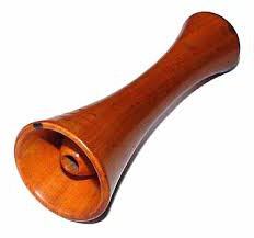
Auscultation (listening) is one of the main methods of clinical examination of a patient, which consists of listening to sound phenomena that spontaneously arise in the body. In practice, both direct or direct auscultation is performed (listening directly with the ear attached to the patient’s body) and indirect auscultation using a stethoscope or phonendoscope. With direct auscultation, audibility is much better than when using a stethoscope, since the auscultated noises are not distorted; for example, weak bronchial breathing , see at aortic insufficiency
For indirect auscultation, solid stethoscopes made of wood, or flexible binaural stethoscopes, consisting of a funnel and two rubber tubes, a stethophonendoscope, and a phonendoscope are used (Fig. 1-3). The stethoscope is a closed acoustic system; it is the main conductor of sound, especially in a flexible stethoscope; in the case of communication with outside air or the lumen of the tube is closed, auscultation becomes almost impossible. When a solid stethoscope is placed into the auricle, the conduction of sound through the bones is of some importance. When auscultating with a stethoscope, noises are more or less distorted due to resonance, but better noise delineation is provided of different origins in a small area (for example, during auscultation of the heart); Noises are perceived more clearly.
Auscultation is of exceptional importance in examining the lungs, heart and blood vessels, measuring blood pressure according to Korotkoff, and determining bowel sounds.
Rice. 1. Plastic stethoscope. Rice. 2. Binaural stethoscope. Rice. 3. Stethoscope.
Auscultation (lat. auscultatio - listening) is one of the main methods clinical trial, which consists of listening to sound phenomena that spontaneously arise in the body.
Auscultation was studied and introduced into medical practice by K. Laennec. The nature of acoustic phenomena detected during auscultation depends on physical properties(density, stress, mass) of oscillating bodies, anatomical structure and intensity of organ function. Oscillations tissue structures occur during the act of breathing, contraction of the heart, movements of the stomach and intestines; Some of the vibrations reach the surface of the body (skin). Each point on the surface of the body is a source of sound waves propagating in all directions; As you move away from the sound source, the wave energy is distributed over increasingly larger volumes of air, so the amplitude of vibrations quickly decreases, and the sound becomes so quiet that it is not perceived by the ear that is not in contact with the body. The main significance of the stethoscope, as well as the direct application of the ear to the body, is that it prevents the sound from being attenuated by dissipating energy.
All sounds occurring in the body are noises, that is, a mixture of sounds of different frequencies. The ear is most sensitive to sounds around 2000 Hz. As frequency decreases, sensitivity decreases sharply, so that at the same intensity, high-frequency sounds seem louder than low-frequency sounds. The ear can more easily detect changes in frequency or pitch than in sound intensity. A weak sound after a strong one is difficult to perceive; In addition, in a mixture of sounds of different frequencies, strong vibrations of one frequency mask weaker vibrations of other frequencies.
The sounds perceived during auscultation of the lungs and heart do not exceed 1000 Hz in frequency. The ear only picks up about 10% of the vibrations caused by the heart ( normal heart is a source of oscillations with a frequency of 5 to 800 per 1 sec.), since some of the oscillations are too low frequency (below 20), i.e. below the threshold of perception (infrasounds), and some are of low intensity. Nevertheless, the importance of auscultation is very great. Phonocardiographic studies have confirmed the explanations for almost all sound phenomena of the heart (tones, noises) established by clinical auscultation (see Phonography).
In practice, both direct or direct auscultation is used, as well as indirect auscultation using a stethoscope, phonendoscope, etc. With direct auscultation, audibility is much better than when using a stethoscope, since the auscultated noises are perceived directly by the auricle and are not distorted. For example, weak bronchial breathing and diastolic murmur in aortic insufficiency are sometimes detected only with this method. V.P. Obraztsov proposed an original method of direct auscultation for recognizing an additional tone during a gallop rhythm. A necessary condition This method of auscultation involves tightly pressing the ear to the area of the heart, in which a closed air cavity is formed and the first heart sound and gallop rhythm acquire the character of a ringing sound due to the push of the heart against the chest, since the gallop rhythm is a tone-push.
For indirect auscultation, solid stethoscopes made of wood, plastic or flexible binaural stethoscopes, consisting of a funnel and two rubber tubes, stethophonendoscopes and phonendoscopes are used.
The stethoscope is a closed acoustic system, and the air in it is the main conductor of sound, especially in a flexible stethoscope; in the case of communication with outside air or the lumen of the tube is closed, auscultation becomes almost impossible. When the solid stethoscope is pointed towards auricle The conduction of sound through the bones of the skull is of some importance. When auscultating with a stethoscope, noises are more or less distorted due to resonance, but better localization and delimitation of noises of different origins in a small area is ensured (for example, during auscultation of the heart); Noises are perceived more clearly. A necessary condition for successful auscultation is silence in the room or office.
Auscultation is an indispensable method of clinical research. It is of exceptional importance in examining the lungs, heart and blood vessels, measuring Korotkoff blood pressure, determining bowel sounds, examining joints, etc.