Show how the human heart works. Heart, its structure and work
The main task of the human heart is to create and maintain a difference in blood pressure in the arteries and veins. It is the difference in pressure that underlies the movement of blood. When the heart stops, blood circulation automatically levels out and stops, thus death occurs. To keep blood moving through the arteries and veins, the body uses many functions of the heart. What role each function plays will be discussed in today’s review.
Before considering the functions of the cardiovascular system, the structure of the heart should be briefly discussed.
The heart in its structure has cavities and chambers consisting of atria and ventricles, which are separated by a septum. Due to the latter, venous and aortic blood does not mix. The atrium and ventricle of each cavity communicate with each other through valves. The chambers are lined with endocardium, and their folds create valves.
Venous blood, saturated with carbon dioxide, collects in the vena cava, which originates in the right atrium. Next, it goes to the right ventricle. Arterial blood is formed in the pulmonary trunk and delivered to the lungs. Blood moves into the left chamber: the atrium and the left ventricle.
Valves play an important role in pumping blood because... like pumps. Automatic operation of the valves allows maintaining blood pressure. During normal heart function, the frequency of its contractions is, on average, 70 beats per minute. It is worth noting that the work of the organ departments - the atria and ventricles - is performed in a sequential manner.
Contraction of the heart muscle is called systolic function, and relaxation is called diastolic function.
The heart muscle or myocardium is the basis of the mass of the organ. The myocardium has a complex structure in the form of layers. The thickness in each part of the human heart can vary from 6 to 11 mm. This muscle works due to electrical impulses, the conductivity of which is ensured by the organ in an independent mode. It is these signals that encourage the heart to work automatically. Outside, the organ is located in a membrane (pericardium), which consists of 2 layers - external and internal (epicardium). In the space between the layers there is 15 ml of serous fluid, due to which sliding occurs during contraction and relaxation.

A brief overview of the structure of the main organ of the human body allows us to talk about the functions of the heart, which are:
- Automaticity - the production of electrical signals even in the absence of external irritation.
- Conduction – excitation of the fibers of the heart and myocardium.
- Excitability is the ability of cells and myocardium to be irritated under the influence of external factors.
- Contractility is the ability of the heart muscle to contract and relax.
The combined concept of the above functions is the autowave function. The pumping function of the heart is ensured and maintained due to the activity of the organ. But in addition to the main task, the heart also performs secondary ones - pumping and endocrine. These functions will be discussed in detail below.
Pressure function
Blood is pumped into the vessels due to periodic contraction of the heart cells of the muscles of the atria and stomachs. The myocardium, contracting, creates high pressure and pushes blood out of the chambers. Due to the fact that the myocardium has a layered structure, the right and left atria and ventricles receive an impulse to contract (automatically) and then relax the muscle. This is called the heart rate. Due to it, the heart fills with blood, carrying it to other organs.

The pumping function of the heart is due to several reasons:
- Based on the remaining inert force caused by the previous contraction of the muscle walls.
- Muscle contraction, which causes compression of the veins in the limbs. Each vein has valves that direct blood along only one vector of movement, i.e. to the heart. Systematic compression ensures blood is pumped to the organ.
- Blood flow to the organ due to inhalation and exhalation of the chest cavity. As a person inhales, the vena cava in the chest stretches and the pressure in the atria becomes low. Therefore, the blood begins to move stronger towards the heart.
Due to the pumping function, the human heart has different pressures in the vessels and moves in one direction due to the valve system.
Endocrine function
The endocrine function of the heart in modern medicine has received a new name – neuroendocrine. This function is responsible for the regulation and coordination of all systems and organs of the human body. The endocrine system adapts the body to constant changes occurring both in the external and internal environment. The result of normal operation of the system is the preservation of homeostasis (author’s note - maintaining balance in the work of all organs and systems).
Based on research conducted in recent years, doctors have identified two new factors:
- The endocrine function of the heart directly interacts with the immune system.
- The heart is the main endocrine gland.

In turn, other systems provide endocrine function:
- glands and hormones;
- transport route;
- tissues and organs that are provided with normal receptor mechanisms.
In other words, this system is aimed at maintaining stability within the body. In addition, the endocrine function, together with human immunity and the central nervous system, ensures reproductive functions, and is also responsible for the growth of new cells and the disposal of “internal waste”.
Based on this, it should be noted that all systems of the human body, brought by nature to automaticity, allow the heart to beat and maintain life.
Pumping function
The cardiac cycle occurs from one muscle contraction to the next. A contraction is created due to the excitation of the myocardium by the heart’s own impulse (function of automatism). This excitation (irritation) is gradually transmitted to the atria and causes a systolic state (author's note - blood pressure). The reaction is then transmitted to the ventricles, causing a systolic state and squeezing blood into the aorta and pulmonary arteries. After this release, the walls of the myocardium relax, the pressure level decreases, and the main organ prepares for the next impulse. Thus, the pumping function of the heart occurs.
Right and left ventricles of the heart

The ventricles are responsible for the hemodynamic task of the human heart. This occurs due to consistent and rhythmic contractions of the left and right atria and ventricles in an automatic mode, which alternate with a state of relaxation of the muscle walls.
The ventricle of the right atrium is located in the front of the human heart and occupies it almost completely. Its structure has denser walls, because Unlike the left ventricle, it contains three layers of myocardium. Based on this, the right ventricle has three sections: inlet, outlet and muscular section. The inner part of the muscular section has a smooth surface, but on the side of the wall there are fleshy crossbars (trabeculae), which are the beginning of the papillary muscles: anterior, posterior and septal. In medical practice, cases have been recorded when there were more of these muscles.
The left ventricle is located in the posterior lower part of the heart. This ventricle is smaller than the right one. But in structure they have minor differences, which are as follows:
- the walls are thinner, due to the presence of only 2 layers of myocardium;
- weakly defined septum.
Despite the slight differences, the functions of the ventricles of the heart are different. Scientists have not yet been able to fully study the chambers of the heart, but the forecast that the main organ is able to very quickly adapt to overload has already received worldwide recognition.
Speaking about the hemodynamic function of the stomachs, it should be noted. The right stomach is the chamber of the organ from which blood circulation begins, directed in a small circle. And the left ventricle is presented in the form of one of the chambers and is the source for the systemic circulation. The left ventricle carries out uninterrupted conduction of blood throughout the body.
The shape of the heart is not the same from person to person. It is determined by age, gender, physique, health, and other factors. In simplified models, it is described by a sphere, ellipsoids, and the intersection figures of an elliptical paraboloid and a triaxial ellipsoid. The measure of elongation (factor) of the shape is the ratio of the largest longitudinal and transverse linear dimensions of the heart. With a hypersthenic body type, the ratio is close to one, and with an asthenic body type, it is about 1.5. The length of the heart of an adult varies from 10 to 15 cm (usually 12-13 cm), width at the base 8-11 cm (usually 9-10 cm) and anteroposterior size 6-8.5 cm (usually 6.5-7 cm) . The average heart weight in men is 332 g (from 274 to 385 g), in women - 253 g (from 203 to 302 g).
In relation to the midline of the body, the heart is located asymmetrically - about 2/3 to the left of it and about 1/3 to the right. Depending on the direction of the projection of the longitudinal axis (from the middle of its base to the apex) on the anterior chest wall, transverse, oblique and vertical positions of the heart are distinguished. The vertical position is more common in people with a narrow and long chest, the transverse position is more common in people with a wide and short chest. The heart can independently provide venous return only in the vessels currently located above the top of the atria, that is, by gravity, by gravity. Performing pumping functions in the circulatory system, the heart constantly pumps blood into the arteries. Simple calculations show that over the course of 70 years, the heart of an average person performs more than 2.5 billion beats and pumps 250 million liters of blood.
Structure of the heart
The heart is located on the left side of the chest in the so-called pericardium, which separates the heart from other organs. The heart wall consists of three layers - the epicardium, myocardium and endocardium. The epicardium consists of a thin (no more than 0.3-0.4 mm) plate of connective tissue, the endocardium consists of epithelial tissue, and the myocardium consists of cardiac striated muscle tissue.
The heart consists of four separate cavities called chambers: left atrium, right atrium, left ventricle, right ventricle. They are separated by partitions. The right atrium contains the hollow veins, and the left atrium contains the pulmonary veins. From the right ventricle and left ventricle emerge, respectively, the pulmonary artery (pulmonary trunk) and the ascending aorta. The right ventricle and left atrium close the pulmonary circulation, the left ventricle and right atrium close the systemic circle. The heart is located in the lower part of the anterior mediastinum, most of its anterior surface is covered by the lungs with the inflowing sections of the vena cava and pulmonary veins, as well as the outflowing aorta and pulmonary trunk. The pericardial cavity contains a small amount of serous fluid.
The wall of the left ventricle is approximately three times thicker than the wall of the right ventricle, since the left must be strong enough to push blood into the systemic circulation for the entire body (the resistance of blood in the systemic circulation is several times greater, and the blood pressure is several times greater higher than in the pulmonary circulation).
There is a need to maintain blood flow in one direction, otherwise the heart could fill with the same blood that was previously sent into the arteries. Responsible for the flow of blood in one direction are the valves, which at the appropriate moment open and close, allowing blood to pass through or blocking it. The valve between the left atrium and the left ventricle is called the mitral valve or bicuspid valve because it consists of two leaflets. The valve between the right atrium and the right ventricle is called the tricuspid valve - it consists of three petals. The heart also contains the aortic and pulmonary valves. They control the flow of blood from both ventricles.
Circulation
Coronary circulation
Each cell of the heart muscle must have a constant supply of oxygen and nutrients. The heart’s own blood circulation, that is, the coronary circulation, is responsible for this process. The name comes from 2 arteries, which, like a crown, entwine the heart. The coronary arteries arise directly from the aorta. Up to 20% of the blood ejected by the heart passes through the coronary system. Only such a powerful portion of oxygenated blood ensures the continuous operation of the life-giving pump of the human body.
Heart cycle
Work of the heart
A healthy heart contracts and unclenches rhythmically and without interruption. There are three phases in one cardiac cycle:
- The atria, filled with blood, contract. In this case, blood is pumped through the open valves into the ventricles of the heart (at this time they remain in a state of relaxation). Contraction of the atria begins at the point where the veins flow into it, so their mouths are compressed and blood cannot flow back into the veins.
- Contraction of the ventricles occurs with simultaneous relaxation of the atria. The tricuspid and bicuspid valves that separate the atria from the ventricles rise, slam shut, and prevent blood from returning to the atria, while the aortic and pulmonary valves open. Contraction of the ventricles forces blood into the aorta and pulmonary artery.
- A pause (diastole) is a relaxation of the entire heart, or a short period of rest for this organ. During a pause, blood from the veins enters the atria and partially flows into the ventricles. When a new cycle begins, the blood remaining in the atria will be pushed into the ventricles - the cycle will repeat.
One cycle of the heart lasts about 0.85 seconds, of which the time of contraction of the atria is only 0.11 seconds, the time of contraction of the ventricles is 0.32 seconds, and the longest is the rest period, lasting 0.4 seconds. The heart of an adult at rest works in the system at about 70 cycles per minute.
Automaticity of the heart
A certain part of the heart muscle specializes in issuing control signals to the rest of the heart in the form of appropriate electrical impulses. These parts of muscle tissue are called the excitatory conduction system. Its main part is the sinoatrial node, called the pacemaker, located on the vault of the right atrium. It controls the heart rate by sending regular electrical impulses. The electrical impulse travels through pathways in the atrium muscle to the atriogastric node. The excited node sends an impulse further to individual muscle cells, causing them to contract. The excitatory conduction system ensures the rhythmic functioning of the heart through synchronized contraction of the atria and ventricles.
Regulation of the heart
The work of the heart is regulated by the nervous and endocrine systems, as well as by Ca and K ions contained in the blood. The work of the nervous system over the heart is to regulate the frequency and strength of heart contractions (the sympathetic nervous system causes increased contractions, the parasympathetic nervous system weakens them). The work of the endocrine system on the heart is to release hormones that increase or decrease heart contractions. The main gland that secretes hormones that regulate the functioning of the heart is the adrenal gland. They secrete the hormones adrenaline and acetylcholine, whose functions in relation to the heart correspond to the functions of the sympathetic and parasympathetic systems. The same work is performed by Ca and K ions, respectively.
Electrical and acoustic phenomena
When the heart (like any muscle) works, electrical phenomena occur that cause the appearance of an electromagnetic field around the working organ. The electrical activity of the heart can be recorded using special electrodes placed on certain areas of the body. Using an electrocardiograph, an electrocardiogram (ECG) is obtained - a picture of changes over time in the potential difference on the surface of the body. ECG plays an important role in diagnosing heart attack and other diseases of the cardiovascular system.
Acoustic phenomena called heart sounds can be heard by placing an ear or stethoscope on the chest. Each cardiac cycle is normally divided into 4 tones. The ear hears the first 2 with each contraction. The longer and lower one is associated with the closure of the bicuspid and tricuspid valves, the shorter and higher one is associated with the closure of the aortic and pulmonary artery valves. Between the first and second tone there is a phase of ventricular contraction.
Notes
see also
Links
| Heart in Wiktionary | |
| Heart on Wikimedia Commons |
Wikimedia Foundation.
2010.
See what “Human Heart” is in other dictionaries:
Wed. I have a lot of silver for feasts, for conversations in red words, for fun of wine. Koltsov. Song. Wed. I didn’t drink beer before I retired: Ask, the whole block will tell you. Now, out of grief, when I get drunk, it’s as if I’m having fun. A.E. Izmailov. Drunkard. Wed. Wine... ...
Wine gladdens the human heart. Wed. I have a lot of silver for feasts, for conversations of red words, for fun of wine. Koltsov. Song. Wed. I didn’t drink before I retired: Ask, the whole block will tell you. Now I get drunk out of grief, It’s as if... ... The womb of a wolf is insatiable, and the heart of a man is insatiable. Wed. Is it enough? “Not yet!” It wouldn't crack. "Don't be afraid." Look, you have become Croesus. “A little more, a little more: At least throw in a handful.” Hey, that's enough! Look, the bag is already crawling apart. “One more pinch!” But here... ...
Michelson's Large Explanatory and Phraseological Dictionary (original spelling) Wed. Is it enough? Not yet! It wouldn't crack. Don't be afraid. Look, you have become Croesus. Another, a little more: At least throw in a handful. Hey, that's enough! Look, the sum is already crawling apart. One more pinch! But then the wallet broke through... Krylov. Fortune and the Beggar. Wed. Or it burns... ...
Michelson's Large Explanatory and Phraseological Dictionary
In order to ensure adequate nutrition of internal organs, the heart pumps an average of seven tons of blood per day. Its size is equal to a clenched fist. Throughout life, this organ makes approximately 2.55 billion beats. The final formation of the heart occurs by the 10th week of intrauterine development. After birth, the type of hemodynamics changes dramatically - from feeding on the mother’s placenta to independent, pulmonary breathing.
Muscle fibers (myocardium) are the predominant type of heart cells. They make up its bulk and are located in the middle layer.
The heart has the following structure:
- three membranes - endocardium, myocardium, epicardium;
- pericardial sac;
- chambers with arterial blood - left atrium (LA) and ventricle (LV);
- sections with venous blood - right atrium (RA) and ventricle (RV);
- valves between the LA and LV (mitral) and tricuspid on the right;
- two valves separate the ventricles and large vessels (aortic on the left and pulmonary artery on the right);
- the septum divides the heart into right and left halves;
- efferent vessels, arteries - pulmonary (venous blood from the pancreas), aorta (arterial from the left ventricle);
- afferent veins - pulmonary (with arterial blood) enter the LA, vena cava flow into the RA.
Internal anatomy and structural features of valves, atria, ventricles
Each part of the heart has its own function and anatomical features. In general, the LV is more powerful (compared to the right), as it forces blood into the arteries, overcoming the high resistance of the vascular walls. The PP is more developed than the left, it receives blood from the whole body, and the left one only from the lungs.
Which side of a person's heart is on?
In humans, the heart is located on the left side in the center of the chest. The main part is located in this area - 75% of the total volume. One third extends beyond the midline into the right half. In this case, the axis of the heart is inclined (oblique direction). This situation is considered classic, as it occurs in the vast majority of adults. But options are also possible:
- dextrocardia (right side);
- almost horizontal - with a wide, short chest;
- close to vertical - for thin people.
Where is a person's heart located?
The human heart is located in the chest between the lungs. It is adjacent to the sternum from the inside, and is limited below by the diaphragm. It is surrounded by a pericardial sac called the pericardium. Pain in the heart area appears on the left near the mammary gland. The top is projected there. But with angina, patients feel pain behind the sternum, and it spreads along the left side of the chest.
Where is the heart located in the human body?
The heart in the human body is located in the center of the chest, but its main part goes into the left half, and only one third is located on the right side. For most people it has an angle of inclination, but for overweight people its position is closer to horizontal, and for thin people it is closer to vertical.
Location of the heart in the human chest
In humans, the heart is located in the chest in such a way that its anterior and lateral surfaces are in contact with the lungs, and its posterior and inferior surfaces are in contact with the diaphragm. The base of the heart (from above) passes into large vessels - the aorta, pulmonary artery. The apex is the lowest part, it approximately corresponds to the 4-5 space between the ribs. It can be found in this area by lowering an imaginary perpendicular from the center of the left collarbone.
The external structure of the heart refers to its chambers; it contains two atria and two ventricles. They are separated by partitions. The pulmonary veins, the vena cava, enter the heart, and the arteries of the lungs, the aorta, carry the blood out. Between the large vessels, at the border of the atria and ventricles of the same name, there are valves:
- aortic;
- pulmonary artery;
- mitral (left);
- tricuspid (between the right parts).
The heart is surrounded by a cavity containing a small amount of fluid. It is formed by the pericardial layers.
If you clench your fist, you can imagine exactly the appearance of a heart. In this case, the part that is located at the wrist joint will be its base, and the acute angle between the first and thumb will be its apex. What is important is that its size is also very close to a clenched fist.
 This is what a human heart looks like
 Borders of the heart and their projection onto the surface of the chest
The boundaries of the heart are found by percussion, by tapping; radiography or echocardiography helps to determine them more accurately. The projections of the cardiac contour onto the surface of the chest are:
- right – 10 mm to the right of the sternum;
- left – 2 cm inward from the perpendicular from the center of the collarbone;
- apex – 5th intercostal space;
- base (upper) – 3rd rib.
What tissues make up the heart?
The heart consists of the following types of tissue:
- muscle - the main one, is called the myocardium, and the cells are cardiomyocytes;
- connective – valves, chords (threads that hold the valves), outer (epicardial) layer;
- epithelium – inner lining (endocardium).

Surfaces of the human heart
The human heart has the following surfaces:
- ribs, sternum – anterior;
- pulmonary – lateral;
- diaphragmatic – lower.
Apex and base of the heart
The apex of the heart is directed down and to the left, its localization is the 5th intercostal space. It represents the top of the cone. The wide part (base) is located on top, closer to the collarbones, and is projected at the level of the 3rd rib.
Human heart shape
The shape of a healthy person's heart is like a cone. Its tip is directed at an acute angle down and to the left of the center of the sternum. The base contains the mouths of large vessels and is located at the level of the 3rd rib.
Right atrium
Receives blood from the vena cava. Next to them is the foramen ovale, which connects the RA and LA in the fetal heart. In a newborn, it closes after the pulmonary blood flow opens, and then completely heals. During systole (contraction), venous blood passes into the pancreas through the tricuspid valve. The RA has a fairly powerful myocardium and a cubic shape.
Left atrium
Arterial blood from the lungs passes into the LA through 4 pulmonary veins and then flows through the opening into the LV. The walls of the LA are 2 times thinner than those of the right one. The shape of the LP is similar to a cylinder.
Right ventricle
It looks like an inverted pyramid. The capacity of the pancreas is about 210 ml. It can be divided into two parts - the arterial (pulmonary) cone and the ventricular cavity itself. In the upper part there are two valves: the tricuspid and the pulmonary trunk.
Left ventricle
It looks like an inverted cone, its lower part forms the top of the heart. The thickness of the myocardium is the largest - 12 mm. There are two openings at the top - for connection with the aorta and LA. Both of them are covered by valves - the aortic and mitral.
Why are the walls of the atria thinner than the walls of the ventricles?
The thickness of the walls of the atrium is less, they are thinner, since they only need to push blood into the ventricles. The right ventricle follows them in strength; it throws its contents into the neighboring lungs, and the left one is the largest in terms of the size of its walls. It pumps blood to the aorta, where there is high pressure.
Tricuspid valve
The right atrioventricular valve consists of a sealed ring that limits the opening and leaflets; there may be not 3, but from 2 to 6.
Half of the people have a tricuspid configuration.
The function of this valve is to prevent the reflux of blood into the RA during RV systole.
Pulmonary valve
It prevents blood from passing back into the pancreas after it contracts. The composition contains valves similar in shape to a crescent. In the middle of each there is a knot that seals the closure.
Mitral valve
It has two doors, one is in the front and the other is in the back. When the valve is open, blood flows from the LA to the LV. When the ventricle contracts, its parts close together to allow blood to pass into the aorta.
Aortic valve
Formed by three semilunar-shaped flaps. Like the pulmonary one, it does not contain threads that hold the valves in place. In the area where the valve is located, the aorta expands and has depressions called sinuses.
Adult heart weight
Depending on the physique and total body weight, the weight of the heart in an adult varies from 200 to 330 g. In men, it is on average 30-50 g heavier than in women.
Diagram of blood circulation
Gas exchange occurs in the alveoli of the lungs. They receive venous blood from the pulmonary artery emerging from the pancreas.
Despite the name, the pulmonary arteries carry venous blood. After the release of carbon dioxide and oxygen saturation through the pulmonary veins, the blood passes into the left atrium. This is how a small circle of blood flow, called pulmonary, is formed.
The large circle covers the entire body as a whole. From the LV, arterial blood spreads to all vessels, nourishing the tissues. Deprived of oxygen, venous blood flows from the vena cava into the RA, then into the RV. The circles close together, ensuring a continuous flow. In order for blood to enter the myocardium, it must first pass into the aorta and then into the two coronary arteries.
They are named so because of the shape of the branches, reminiscent of a crown (crown). Venous blood from the heart muscle predominantly enters the coronary sinus. It opens into the right atrium. This circle of blood circulation is considered the third, coronary.
Watch the video about the structure of the human heart:
What is special about the structure of a child’s heart?
Until the age of six, the heart is spherical due to the large atria. Its walls are easily stretched, they are much thinner than those of adults. A network of tendon threads is gradually formed, fixing the valve leaflets and papillary muscles. Full development of all heart structures ends by age 20.
The position of the newborn's heart in the chest is initially oblique, adjacent to the anterior surface. This is caused by an increase in the volume of lung tissue and a decrease in the mass of the thymus gland.
Until two years of age, the heart impulse forms the right ventricle, and then part of the left. The atria are the leaders in growth rate up to 2 years, and the ventricles after 10 years. Up to ten years, the LV is ahead of the right.
Basic functions of the myocardium
- The heart muscle differs in structure from all others, as it has several unique properties:
- Automatism is excitation under the influence of one’s own bioelectric impulses. They first form in the sinus node. He is the main pacemaker, generating about 60 - 80 signals per minute. The underlying cells of the conduction system are nodes of the 2nd and 3rd order.
- Excitability - in response to external and internal stimuli, the myocardium is activated.
- Contractility is the ability to contract when excited. This function creates the pumping capabilities of the heart. The force with which the myocardium reacts to an electrical stimulus depends on the pressure in the aorta, the degree of stretching of the fibers in diastole, and the volume of blood in the chambers.
The functioning of the heart goes through three stages:
- Contraction of the RA, LA and relaxation of the RV and LV with the opening of the valves between them. Transition of blood into the ventricles.
- Ventricular systole - the valves of the blood vessels open, blood flows into the aorta and pulmonary artery.
- General relaxation (diastole) - blood fills the atria and presses on the valves (mitral and tricuspid) until they open.
During the period of contraction of the ventricles, the valves between them and the atria are closed by blood pressure. In diastole, the pressure in the ventricles drops, it becomes lower than in large vessels, then parts of the pulmonary and aortic valves close so that the blood flow does not return.
Heart cycle
There are 2 stages in the heart cycle: contraction and relaxation. The first is called systole and also includes 2 phases:
- compression of the atria to fill the ventricles (lasts 0.1 sec.);
- the work of the ventricular part and the release of blood into large vessels (about 0.5 sec.).
Then comes relaxation - diastole (0.36 sec). Cells change polarity to respond to the next impulse (repolarization), and the blood vessels of the myocardium bring nutrition. During this period, the atria begin to fill.
The heart ensures the movement of blood through the large and small circles thanks to the coordinated work of the atria, ventricles, great vessels and valves.
The myocardium has the ability to generate an electrical impulse and conduct it from the nodes of automaticity to the cells of the ventricles. In response to the signal, muscle fibers become active and contract. The cardiac cycle consists of a systolic and a diastolic period.
Useful video
Watch the video about the work of the human heart:
Read also


© Use of site materials only in agreement with the administration.
The structure of the heart of any organism has many characteristic nuances. In the process of phylogenesis, that is, the evolution of living organisms to more complex ones, the heart of birds, animals and humans acquires four chambers instead of two chambers in fish and three chambers in amphibians. This complex structure is best suited for separating the flow of arterial and venous blood. In addition, the anatomy of the human heart involves many small details, each of which performs its own strictly defined functions.
Heart as an organ
So, the heart is nothing more than a hollow organ consisting of specific muscle tissue, which carries out the motor function. The heart is located in the chest behind the sternum, more to the left, and its longitudinal axis is directed anteriorly, to the left and down. In front, the heart borders on the lungs, almost completely covering them, leaving only a small part directly adjacent to the chest from the inside. The boundaries of this part are otherwise called absolute cardiac dullness, and they can be determined by tapping the chest wall ().
In people with a normal constitution, the heart has a semi-horizontal position in the chest cavity, in people with an asthenic constitution (thin and tall) it is almost vertical, and in hypersthenics (dense, stocky, with large muscle mass) it is almost horizontal.

heart position
The posterior wall of the heart is adjacent to the esophagus and to the large main vessels (thoracic aorta, inferior vena cava). The lower part of the heart is located on the diaphragm.

external structure of the heart
Age characteristics
The human heart begins to form in the third week of the intrauterine period and continues throughout the entire period of gestation, passing through stages from a single-chamber cavity to a four-chamber heart.

development of the heart in utero
The formation of four chambers (two atria and two ventricles) occurs already in the first two months of pregnancy. The smallest structures are fully formed by birth. It is in the first two months that the heart of the embryo is most vulnerable to the negative influence of certain factors on the expectant mother.
The fetal heart participates in the blood flow throughout its body, but it differs in the circles of blood circulation - the fetus does not yet have its own breathing with its lungs, but “breathes” through the placental blood. There are some openings in the fetal heart that allow pulmonary blood flow to be “switched off” from the circulation before birth. During childbirth, accompanied by the first cry of the newborn, and, consequently, at the moment of increased intrathoracic pressure and pressure in the baby's heart, these openings close. But this does not always happen, and the child may still have them, for example (not to be confused with a defect such as atrial septal defect). An open window is not a heart defect, and subsequently, as the child grows, it closes.
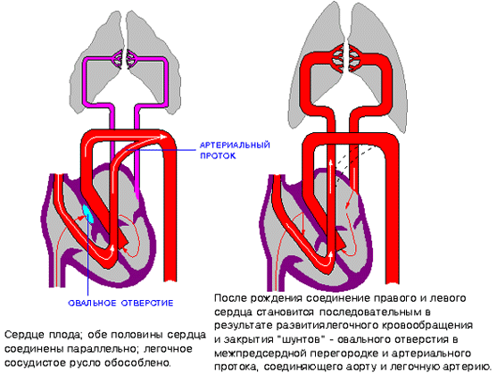
hemodynamics in the heart before and after birth
The heart of a newborn baby has a round shape, and its dimensions are 3-4 cm in length and 3-3.5 cm in width. In the first year of a child's life, the heart increases significantly in size, more in length than in width. The weight of a newborn baby's heart is about 25-30 grams.
As the baby grows and develops, the heart also grows, sometimes significantly ahead of the development of the body itself according to age. By the age of 15, the mass of the heart increases almost tenfold, and its volume increases more than fivefold. The heart grows most rapidly until the age of five, and then during puberty.
In an adult, the size of the heart is about 11-14 cm in length and 8-10 cm in width. Many people rightly believe that the size of each person’s heart corresponds to the size of his clenched fist. The weight of the heart in women is about 200 grams, and in men it is about 300-350 grams.
After age 25, changes begin in the connective tissue of the heart, which forms the heart valves. Their elasticity is no longer the same as in childhood and adolescence, and the edges may become uneven. As a person grows and then ages, changes occur in all structures of the heart, as well as in the vessels that feed it (the coronary arteries). These changes can lead to the development of numerous cardiac diseases.
Anatomical and functional features of the heart
Anatomically, the heart is an organ divided into four chambers by septa and valves. The “upper” two are called atria (atrium), and the “lower” two are called ventricles (ventriculum). Between the right and left atria is the interatrial septum, and between the ventricles is the interventricular septum. Normally, these septa do not have holes in them. If there are holes, this leads to mixing of arterial and venous blood, and, accordingly, to hypoxia of many organs and tissues. Such holes are called septal defects and are classified as.

basic structure of the chambers of the heart
The boundaries between the upper and lower chambers are the atrioventricular openings - the left one, covered by the mitral valve leaflets, and the right one, covered by the tricuspid valve leaflets. The integrity of the septa and the proper operation of the valve leaflets prevent the mixing of blood flows in the heart and promote clear unidirectional blood flow.
The atria and ventricles are different - the atria are smaller than the ventricles and have thinner walls. Thus, the wall of the atria is about only three millimeters, the wall of the right ventricle is about 0.5 cm, and the wall of the left is about 1.5 cm.
The atria have small projections called ears. They have a slight suction function for better pumping of blood into the atrium cavity. The mouth of the vena cava flows into the right atrium near its appendage, and four (less often five) pulmonary veins flow into the left atrium. The pulmonary artery (more often called the pulmonary trunk) on the right and the aortic bulb on the left depart from the ventricles.

structure of the heart and its vessels
From the inside, the upper and lower chambers of the heart are also different and have their own characteristics. The surface of the atria is smoother than the ventricles. Thin connective tissue valves originate from the valve ring between the atrium and the ventricle - bicuspid (mitral) on the left and tricuspid (tricuspid) on the right. The other edge of the valves faces the inside of the ventricles. But so that they do not hang freely, they are supported, as it were, by thin tendon threads called chords. They are like springs, stretch when the valve flaps close and compress when the valve flaps open. The chordae originate from the papillary muscles from the wall of the ventricles - three in the right and two in the left ventricle. That is why the ventricular cavity has an uneven and lumpy inner surface.
The functions of the atria and ventricles also differ. Due to the fact that the atria need to push blood into the ventricles, and not into larger and longer vessels, they have to overcome less resistance from muscle tissue, therefore the atria are smaller in size and their walls are thinner than those of the ventricles. The ventricles push blood into the aorta (left) and the pulmonary artery (right). Conventionally, the heart is divided into right and left halves. The right half serves for the flow of exclusively venous blood, and the left half for arterial blood. Schematically, the “right heart” is indicated in blue, and the “left heart” is indicated in red. Normally, these flows never mix.

hemodynamics in the heart
One cardiac cycle lasts about 1 second and is carried out as follows. At the moment the atria are filled with blood, their walls relax - atrial diastole occurs. The valves of the vena cava and pulmonary veins are open. The tricuspid and mitral valves are closed. Then the atrial walls tense and push blood into the ventricles, the tricuspid and mitral valves are open. At this moment, systole (contraction) of the atria and diastole (relaxation) of the ventricles occur. After the ventricles receive blood, the tricuspid and mitral valves close, and the aortic and pulmonary valves open. Next, the ventricles contract (ventricular systole), and the atria fill with blood again. The general diastole of the heart begins.

Cardiac cycle
The main function of the heart is reduced to pumping, that is, to pushing a certain blood volume into the aorta with such pressure and speed that the blood is delivered to the most distant organs and to the smallest cells of the body. Moreover, arterial blood with a high content of oxygen and nutrients is pushed into the aorta, entering the left half of the heart from the vessels of the lungs (flows to the heart through the pulmonary veins).
Venous blood, low in oxygen and other substances, is collected from all cells and organs from the venous cava system, and flows into the right half of the heart from the superior and inferior vena cava. Next, venous blood is pushed from the right ventricle into the pulmonary artery, and then into the pulmonary vessels in order to carry out gas exchange in the alveoli of the lungs and to enrich it with oxygen. In the lungs, arterial blood collects in the pulmonary venules and veins, and again flows into the left side of the heart (the left atrium). And so the heart regularly pumps blood throughout the body at a frequency of 60-80 beats per minute. These processes are designated by the concept "Circles of Blood Circulation". There are two of them - small and large:
- Small circle includes the flow of venous blood from the right atrium through the tricuspid valve into the right ventricle - then into the pulmonary artery - then into the arteries of the lungs - oxygenation of blood in the pulmonary alveoli - flow of arterial blood into the smallest veins of the lungs - into the pulmonary veins - into the left atrium.
- Big circle includes the flow of arterial blood from the left atrium through the mitral valve into the left ventricle - through the aorta into the arterial bed of all organs - after gas exchange in tissues and organs, the blood becomes venous (with a high content of carbon dioxide instead of oxygen) - then into the venous bed of organs - into the hollow system veins - into the right atrium.

circulation circles
Video: cardiac anatomy and cardiac cycle briefly
Morphological features of the heart
If you examine sections of the heart under a microscope, you can see a special type of muscle that is not found in any other organ. This is a type of striated muscle, but has significant histological differences from ordinary skeletal muscles and from the muscles lining internal organs. The main function of the heart muscle, or myocardium, is to provide the most important ability of the heart, which forms the basis for the vital activity of the entire organism as a whole. This is the ability to contract, or contractility.
In order for the heart muscle fibers to contract synchronously, electrical signals must be supplied to them, which excite the fibers. This is another ability of the heart – .
Conduction and contractility are possible due to the fact that the heart autonomously generates electricity. Function data (automatism and excitability) are provided by special fibers that are an integral part of the conductive system. The latter is represented by electrically active cells of the sinus node, atrioventricular node, the bundle of His (with two legs - right and left), as well as Purkinje fibers. In the case when a patient’s myocardial damage affects these fibers, they develop, otherwise called.
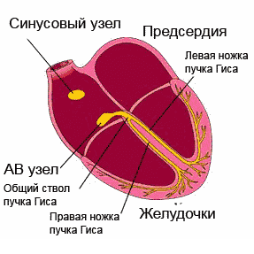
cardiac cycle
Normally, the electrical impulse originates in the cells of the sinus node, which is located in the area of the right atrium appendage. In a short period of time (about half a millisecond), the impulse spreads throughout the atrial myocardium and then enters the cells of the atrioventricular junction. Typically, signals are transmitted to the AV node through three main tracts - the Wenkenbach, Thorel and Bachmann bundles. In the cells of the AV node, the impulse transmission time is extended to 20-80 milliseconds, and then the impulses travel through the right and left legs (as well as the anterior and posterior branches of the left leg) of the His bundle to the Purkinje fibers, and ultimately to the working myocardium. The frequency of impulse transmission along all pathways is equal to the heart rate and is 55-80 impulses per minute.
So, the myocardium, or heart muscle, is the middle layer in the wall of the heart. The inner and outer membranes are connective tissue and are called endocardium and epicardium. The last layer is part of the pericardial sac, or cardiac “shirt”. Between the inner layer of the pericardium and the epicardium, a cavity is formed, filled with a very small amount of fluid, to ensure better sliding of the pericardial layers during heart contractions. Normally, the fluid volume is up to 50 ml; exceeding this volume may indicate pericarditis.

structure of the heart wall and membrane
Blood supply and innervation of the heart
Despite the fact that the heart is a pump to supply the entire body with oxygen and nutrients, it itself also needs arterial blood. In this regard, the entire wall of the heart has a well-developed arterial network, which is represented by the branching of the coronary (coronary) arteries. The orifices of the right and left coronary arteries depart from the root of the aorta and are divided into branches that penetrate into the thickness of the heart wall. If these important arteries become clogged with blood clots and atherosclerotic plaques, the patient will develop and the organ will no longer be able to perform its functions fully.

location of the coronary arteries supplying blood to the heart muscle (myocardium)
The frequency and force with which the heart beats is influenced by nerve fibers extending from the most important nerve conductors - the vagus nerve and the sympathetic trunk. The first fibers have the ability to slow down the rhythm frequency, the latter - to increase the frequency and strength of the heartbeat, that is, they act like adrenaline.

innervation of the heart
In conclusion, it should be noted that the anatomy of the heart may have any deviations in individual patients, therefore, only a doctor can determine the norm or pathology in a person after conducting an examination that can most informatively visualize the cardiovascular system.
Video: lecture on cardiac anatomy
The heart is one of the most perfect organs of the human body, which was created with special thought and care. He has excellent qualities: fantastic power, rare tirelessness and an inimitable ability to adapt to the external environment. It’s not for nothing that many people call the heart the human motor, because in fact, it is so. If you just think about the colossal work of our “engine”, then this is a most amazing organ.
What is the heart and what are its functions?
The heart is a muscular organ that, through rhythmic, repeated contractions, ensures blood flow through the blood vessels.
The main function of the heart is to ensure constant and uninterrupted blood flow throughout the body.. Therefore, the heart is a kind of pump that circulates blood throughout the body, and this is its main function. Thanks to the work of the heart, blood flows to all parts of the body and organs, saturates tissues with nutrients and oxygen, and also saturates the blood itself with oxygen. During physical activity, increasing the speed of movement (running) and under stress, the heart must produce an instant reaction and increase the speed and number of contractions.
We have become familiar with what the heart is and what its functions are, now let’s look at the structure of the heart.
To begin with, it is worth saying that the human heart is located on the left side of the chest. It is important to note that there is a group of unique people in the world whose heart is located not on the left side, as usual, but on the right side; such people, as a rule, have a mirror structure of the body, as a result of which the heart is located in the opposite direction from the usual location side.
The heart consists of four separate chambers (cavities):
- Left atrium;
- Right atrium;
- Left ventricle;
- Right ventricle.
The valves located in the heart are responsible for the flow of blood.. The left atrium contains the pulmonary veins and the right atrium - the hollow veins (superior vena cava and inferior vena cava). The pulmonary trunk and the ascending aorta emerge from the left and right ventricles.
The left ventricle shares with the left atrium mitral valve(bicuspid valve). The right ventricle and right atrium are separated tricuspid valve. Also in the very heart are pulmonary and aortic valves, which are responsible for the flow of blood from the left and right ventricles.

Circulation circles of the heart
As you know, the heart produces 2 types of blood circulation circles - this, in turn, is the systemic circulation and the small one. Systemic circulation originates from the left ventricle and ends in the right atrium.The task of the systemic circulation is to supply blood to all organs of the body, as well as directly to the lungs themselves.
Pulmonary circulation originates from the right ventricle and ends in the left atrium.
As for the pulmonary circulation, it is responsible for gas exchange in the pulmonary alveoli.
Here's a brief summary of what concerns the blood circulation.
What does the heart do?
What is a heart for? As you already understand, the heart produces continuous blood flow throughout the body. A three-hundred-gram ball of muscle, elastic and mobile, is a constantly working suction and discharge pump, the right half of which takes the blood used in the body from the veins and directs it to the lungs for enrichment with oxygen. The blood from the lungs then enters the left side of the heart and, with a certain degree of force, measured by the level of blood pressure, ejects the blood.
Blood circulation during blood circulation occurs approximately 100 thousand times a day, at a distance of over 100 thousand kilometers (this is the total length of the vessels of the human body). Over the course of a year, the number of heartbeats reaches an astronomical value - 34 million. During this time, 3 million liters of blood are pumped. Gigantic work! What amazing reserves are hidden in this biological engine!
Interesting to know: one contraction requires enough energy to lift a 400 g load to a height of one meter. Moreover, a calm heart uses only 15% of all the energy it has. With hard work, this figure increases to 35%.
Unlike skeletal muscles, which can remain at rest for hours, the contractile cells of the myocardium work tirelessly for many years. This gives rise to one important requirement: their air supply must be continuous and optimal. If there are no nutrients and oxygen, the cell dies instantly. It cannot stop and wait for delayed doses of life-giving gas and glucose, since it does not create the reserves necessary for the so-called maneuver. Her life lies in a saving breath of fresh blood.
But can a muscle saturated with blood starve? Yes maybe. The fact is that the myocardium does not feed on the blood that fills its cavities. It is supplied with oxygen and essential nutrients through two “pipelines” that branch from the base of the aorta and crown the muscle like a crown (hence their name “coronary” or “coronary”). They, in turn, form a dense network of capillaries that nourish its own tissue. There are a lot of spare branches here - collaterals that duplicate the main vessels and run parallel with them - something like the branches and ducts of a large river. In addition, the basins of the main “blood rivers” are not separated, but are connected into a single whole thanks to transverse vessels - anastomoses. If something bad happens: a blockage or rupture, the blood will rush along the alternate channel and the loss will be more than compensated for. Thus, nature has provided not only the hidden powers of the pumping mechanism, but also a perfect system of replacement blood supply.
This process, common to all vessels, is especially pathological for the coronary arteries. After all, they are very thin, the largest of them is no wider than the straw through which you drink a cocktail. The peculiarity of blood circulation in the myocardium also plays a role. Oddly enough, blood periodically stops in these intensely circulating arteries. Scientists explain this strangeness as follows. Unlike other vessels, the coronary arteries are exposed to two forces that are opposite to each other: the pulse pressure of the blood flowing through the aorta, and the counter pressure that occurs when the heart muscle contracts and tends to push the blood back to the aorta. When the opposing forces become equal, blood flow stops for a split second. This time is enough for some of the clot-forming material to precipitate from the blood. This is why atherosclerosis of the coronary vessels develops many years before it occurs in other arteries.

Heart diseases
Nowadays, cardiovascular diseases are attacking people at an active pace, especially the elderly. Millions of deaths a year - this is the outcome of heart disease. This means: three out of five patients die directly from heart attacks. Statistics note two alarming facts: the trend of increasing diseases and their rejuvenation.Heart diseases include 3 groups of diseases that affect:
- Heart valves (congenital or acquired heart defects);
- Cardiac vessels;
- Tissues of the membranes of the heart.
Heart failure. This term refers to a disease in which a complex of disorders occurs due to a decrease in myocardial contractility, which is a consequence of the development of stagnant processes. In heart failure, blood stagnates in both the pulmonary and systemic circulation.
Heart defects. With heart defects, defects in the functioning of the valve apparatus can be observed, which can lead to heart failure. Heart defects can be either congenital or acquired.
Heart arythmy. This heart pathology is caused