Uterine puncture: diagnostic value and procedure. Carrying out culdocentesis in gynecology: rationale, technique, complications of the intervention
Puncture is a specific procedure that is used to diagnose pathologies as well as treat internal organs, biological cavities. It is done using special needles and other devices. Before agreeing to such a procedure, it is necessary to take a closer look at what a puncture is, what features it has and how it is performed.
A puncture is a special puncture of the tissues of internal organs, blood vessels, various neoplasms, cavities for collecting fluids for the purpose of diagnosing pathologies. In addition, the use of the procedure in some cases is necessary for the administration of medications. It is used to diagnose liver pathologies, bone marrow, lungs, bone tissue. Basically, in this way they are determined oncological diseases. To clarify the diagnosis, materials are taken directly from the tumor. As for the blood vessels, they are punctured for sampling biological fluid, installation of catheters through which medications. Parenteral nutrition is also performed in the same way.
If in the abdominal, articular or pleural cavity If an inflammatory process is observed, accompanied by an accumulation of fluid or pus, then a puncture is used to remove this pathological content. For example, using this procedure, drains are installed for flushing internal organs and administering medications.
Regarding puncture, this is a mandatory procedure used in anesthesiology, especially during operations on the extremities. It is widespread in gynecology to identify a number of diseases and treat them.
Indications for use of the procedure in gynecology
So, to use a puncture puncture there must be appropriate indications. They do it in order to:
- confirm ectopic pregnancy or female factor infertility;
- determine the presence of uterine or internal organ rupture;
- exclude peritonitis;
- counting the number of oocytes in the ovaries;
- determine the amount and nature of exudate in the organ cavity, tumors;
- diagnose internal endometriosis, as well as other neoplasms of a malignant or benign nature;
- determine the violation menstrual cycle, uterine bleeding unspecified genesis;
- diagnose or exclude developmental abnormalities reproductive organs women;
- collect material to determine the effectiveness of the treatment;
- collect eggs during the IVF procedure.
After the puncture, the patient can go home the next day only if a serious illness is not diagnosed.

Types of puncture in gynecology
There are several types of punctures that are used for the diagnosis and treatment of female diseases:

All of these types of punctures are used in gynecology in difficult cases when diagnosis or treatment by other methods does not give a positive result.
General rules for puncture
Many women are interested in how a puncture is performed. In most cases, it is painless. However, in order for the procedure to take place without complications, as well as for the psychological comfort of the woman, anesthesia or pain relief is necessary. There are other rules for performing a puncture:

- Before the procedure, all instruments, as well as the external genitalia, must be treated with a disinfectant solution. This will avoid additional infection of internal tissues and cavities.
- If the puncture is done through back wall vagina, the movement should be sharp and light. At the same time, care must be taken not to damage the wall of the rectum.
- If there is very thick exudate in the cyst or cavity, which can clog the needle, it is necessary to inject a sterile solution inside.
- Puncture is allowed only in specialized clinics or medical offices.
The procedure is quite complex, so it must be carried out by an experienced specialist with a good reputation.
Possible consequences
At all, exploratory operation is painless, but sometimes the following consequences of puncture can be observed:
- injury to blood vessels or the endometroid layer of the uterus;
- decreased blood pressure (during operations involving serious blood loss);
- in the organ or cavity in which the puncture is performed;
- damage to the rectum (often additional treatment not required);
- general deterioration of health;
- dizziness;
- scanty vaginal discharge;
- stupid painful sensations in the abdominal area;
- incorrect diagnosis (blood in the fluid may appear not as a result of the disease, but due to damage to the vessels located in the periuterine tissue).
Puncture in gynecology is a frequently used tool for diagnosing and treating pathologies reproductive system. It can only be done as prescribed by a doctor in a medical facility.
Posterior fornix puncture The vagina allows access to the pelvic cavity. For various pathologies in abdominal cavity fluid accumulates (exudate, blood and pus), puncture helps to eliminate this fluid or take it for research, that is, this procedure serves not only diagnostic, but also therapeutic purposes. Puncture for diagnostic purposes is performed in a hospital setting. With its help it is revealed excess liquid in the pelvic cavity. The resulting sample is sent to full research to the laboratory. There it is studied by bacteriologists and cytologists. Based on the results of this diagnosis, treatment is prescribed. But let's talk about everything in order.
Indications for puncture
More recently this procedure was carried out exclusively in cases of suspected ovarian apoplexy or ectopic pregnancy. But modern medicine is increasingly using laparoscopy for these purposes, although even today puncture is often performed for diagnosis ectopic pregnancy. But the main indications for its implementation are suspicion of malignancy and inflammatory diseases in the pelvic organs.
Contraindications for puncture
Contraindications include severe heart failure. There are no more direct contraindications. But after full examination the doctor can identify factors that make this type of puncture undesirable.
How to prepare for research?
Basically, no preparation is required, but if there are any peculiarities of the body, the doctor may prescribe certain preparatory measures, naturally, after a full examination. Before the procedure you need to release bladder and carry out basic hygiene measures.
Puncture technique
The first step is to carry out local anesthesia. The external genitalia are treated with iodine solution or alcohol. Then it is carried out puncture of the posterior fornix vagina, that is, a needle attached to a syringe is inserted into it under the control of ultrasonic equipment. It is inserted 2 cm deep, and with its help the amount of liquid required for the study is extracted. When performing a therapeutic puncture, fluid is pumped out from internal cavity. Upon completion of the procedure, the needle is slowly and carefully removed. The resulting sample is sent for cytological and bacteriological examination.
Although seemingly simple, such a puncture is actually a very complex procedure. All actions must be carried out very carefully so as not to damage the tissue next to the organ being examined. During the puncture, an inexperienced doctor may well hit some artery, so the puncture should only be performed by an experienced specialist. Come to our center, we will perform the puncture without complications.
Possible complications of puncture
As we wrote above, most complications after this type of puncture occur due to the inexperience of the doctor. Their appearance is also possible if the patient does not comply with the specialist’s recommendations. But this is more of an exception, which is why complications rarely occur. Main complications:
. The needle enters the parametrium vessel;Infection;
Injury to a vessel of the uterus or vagina;
Intestinal injury. All these complications are very dangerous and can lead to serious consequences, so immediate treatment is required to eliminate them.
Where to perform the puncture?
This procedure is best carried out in our center in Moscow, under the close supervision of experienced specialists. To monitor the puncture, our center uses the most modern ultrasound equipment, thanks to which the risk of complications is reduced to zero. By contacting our center, you will receive high-quality treatment and the most accurate diagnosis.
Abdominal puncture through the posterior vaginal fornix involves inserting a needle into the abdominal cavity through the posterior vaginal fornix.
Indications for puncture of the abdominal cavity through the posterior vaginal fornix: suspicion of interrupted ectopic pregnancy, ovarian apoplexy, intra-abdominal bleeding, abscess of the rectal uterine cavity.
Contraindications: heart failure grades 2 and 3, serious condition.
Special equipment. A needle with a diameter of no more than 2 mm and a length of at least 12 cm. There may be a special device with a needle, which can be attached to the wall of the posterior vaginal fornix using a vacuum cup.
Instruments for puncture of the abdominal cavity through the posterior vaginal fornix: spoon-shaped vaginal speculum, vaginal lift, bullet forceps, puncture needle or puncture device, forceps.
Technique of puncture of the abdominal cavity through the posterior vaginal fornix
Empty the bladder. The external genitalia, vagina and cervix are lubricated with iodonate. After inserting the speculum, the posterior lip of the cervix is pulled anteriorly with forceps. The posterior fornix of the vagina is stretched. A needle is inserted into the center of the stretched arch perpendicular to the surface to a depth of 2 cm. This is often enough to obtain liquid, if there is any there, because with a stretched vault, the pelvic peritoneum is closely adjacent to the vaginal wall. With deeper advancement, the needle may enter the intestine or tumor. When moving, the needle should easily overcome the obstacle. If strong resistance is felt, it means there is an obstacle in its path, most likely the uterus. In this case, you need to change the direction of the needle or abandon the puncture.
Fluid from the abdominal cavity may leak through the needle on its own. The liquid can be aspirated using a syringe.
Diagnostic value of abdominal puncture through the posterior vaginal fornix
This study allows for differential diagnosis between a disrupted ectopic pregnancy and inflammation of the uterine appendages.
Blood in the aspirate indicates the presence of intra-abdominal bleeding (interrupted ectopic pregnancy, ovarian apoplexy, trauma to the abdominal organs). In some cases, the needle may enter a vessel or the uterus: then the same blood is sucked into the syringe as during a vein puncture. If there is intra-abdominal bleeding, the blood is dark, with small clots, and does not clot.
There may be pus in the punctate; if there is exudate in the rectal-uterine cavity, there may be exudate (serous, serous-purulent, serous-hemorrhagic). This is typical for patients with inflammatory diseases uterus and its appendages, complicated by pelvioperitonitis, as well as for other inflammatory processes abdominal organs. The resulting liquid in a sterile test tube is sent for bacteriological examination.
The article was prepared and edited by: surgeonVideo:
Healthy:
Related articles:
- Removal of a pedunculated ovarian cyst Indications: ovarian cyst. Patient position: lying on your back. Under...
- Indications for puncture maxillary sinus: spicy and chronic sinusitis. Technique for puncture of the maxillary sinus: after anesthesia of the mucous membrane...
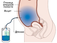
A puncture of the abdominal cavity is performed for the purpose of drainage and examination of fluid that may accumulate in the free space between internal organs or in the pelvic cavity.
The presence of fluid is a symptom of many diseases. To put correct diagnosis, alleviate the patient’s condition and prescribe correct treatment, this is assigned medical procedure. It can be done in two ways. These include culdocentesis and laparocentesis. Culdocentesis is a puncture of the abdominal cavity through the posterior vaginal fornix, performed only in women. The second method, abdominal puncture, is used in both sexes.
Preparation and performance of abdominal puncture
If the patient was prescribed a puncture of the abdominal cavity, so that no complications arise during its implementation and all manipulations performed are positive outcome, you need to prepare properly.
Approximately 2–3 hours before the minimally invasive intervention, the patient needs to undergo a cleansing enema. Immediately before the puncture itself, you should visit the restroom and empty your bladder.
Drainage of the cavity is usually carried out in the manipulation room, that is, an operating room is not required. All devices used during the procedure must be sterile.
A solution of Promedol or Atropine sulfate is used as an anesthetic drug.
If the patient’s condition is serious, then the process of collecting biological material is carried out in a lying position (on the right side). Under other circumstances, the patient is seated in a chair so that he can lean on the back.
The area where the puncture will be performed is treated with a disinfectant. To ensure that the entire process takes place under strict medical supervision, it is carried out using an ultrasound machine. Otherwise, there is a risk of damage to internal organs, which can lead to serious bleeding.
Puncture abdominal wall followed by collection of biological material for analysis, as a rule, using a device such as a trocar. As soon as the liquid begins to come out, its first portions are collected in a pre-prepared sterile container and sent to the laboratory. When a puncture is performed not only for diagnostic purposes, but also to pump out all available fluid, that is, for therapeutic purposes, after collecting biological material for research, pumping out the contents of the abdominal cavity continues. It is collected in a special tank. In 1 session, you can pump out up to 6 liters of liquid. To compensate for the loss of salts and proteins, the patient must be administered a solution of Albumin or its analogues.

The final stage of puncture is the removal of all instruments used and the application surgical sutures. The stitched puncture site is covered with a sterile napkin and bandaged.
When all manipulations are completed, the patient remains under medical supervision. Medical staff monitors:
- blood pressure indicators;
- condition of the skin;
- condition of the mucous membranes;
- general well-being.
Puncture through the posterior vaginal fornix
In gynecology, puncture is used both as therapy and for diagnosis. It may be prescribed if an ectopic pregnancy is suspected or if there are symptoms of an abscess in the pelvic cavity. The puncture is performed using local anesthesia.
Accumulated biological material in the area where the pelvic organs are located may consist of:
- exudate;
- blood;
- pus.
The collected cavity contents are immediately sent for laboratory analysis.
Below the cervix in the area of the posterior fornix between the divergent uterosacral ligaments, the peritoneum comes very close to the walls of the vagina. It is this place that is most convenient for performing a puncture.
After completing the disinfection of the external genitalia, the doctor begins to perform a puncture. Using a speculum, he exposes the vaginal part of the cervix. Special gynecological forceps are used to grasp and bend the posterior lip of the uterus. This is how the posterior arch is stretched.
The puncture needle should enter between the uterosacral ligaments. It is deepened by approximately 2 cm. When the end of the needle is at the required depth, biological material is collected using the syringe plunger.
Although it is required laboratory test, experienced specialist in appearance fluid can make an assumption about what kind of fluid is developing pathological process. For example, liquid blood that has a dark color is characteristic of the termination of an ectopic pregnancy. IN biological material small clots may be seen.
Puncture through the posterior vaginal fornix should be performed by a qualified specialist to eliminate the possibility of obtaining a false positive result and not further harm the patient.
IN Lately puncture through the posterior vaginal fornix is rarely performed, since during recovery period There is big risk addition of a secondary infection. Less traumatic and equally informative is laparoscopic examination. It is preferred because, according to statistics, the risk of complications after this manipulation is minimal.
IN modern medicine there are many most different ways diagnostics that can quickly and accurately confirm or refute the suspected diagnosis. Some techniques are simple and do not require any special preparation from patients. Moreover, the procedures themselves are carried out quickly, without any effort. Other methods can cause discomfort, but it is impossible to do without them. One of these manipulations is puncture of the posterior vaginal fornix.
Features of the procedure
Puncture of the posterior vaginal vault has its own characteristics. It is carried out for diagnostic purposes to identify the contents of the rectouterine cavity. Less commonly, this procedure is performed as an auxiliary procedure.
Puncture of the posterior vaginal fornix requires anesthesia. Patients are given short-term anesthesia or local conduction anesthesia.
For diagnosis to be effective, the patient must lie down so that her pelvis is downward. This position helps to drain even a small amount of fluid located in the rectal-uterine area. This greatly increases the effectiveness of the manipulation.
Indications
Puncture of the posterior vaginal fornix is used in case of suspicion of other internal organs, as well as:
- if you suspect the presence of any type of fluid in the pelvis;
- administer medications if necessary;
- if you suspect ovarian cancer;
- upon breakthrough purulent pathologies into the abdominal cavity.
Puncture through the posterior vaginal fornix allows you to accurately determine the presence of fluid and its type without surgical intervention.

Where is the procedure performed?
The manipulation is carried out only in a hospital, since it is surgical appearance interventions. During the procedure, all rules of antiseptics and asepsis are observed. Before the puncture is performed, the patient must empty her bladder and bowels. For these purposes, a cleansing enema may be prescribed.
Most often, nitrous oxide or any other mask anesthesia is used for anesthesia. Less commonly used general intravenous anesthesia And local anesthesia in the form of a solution of novocaine.
How is it carried out?
Puncture of the abdominal cavity through the posterior vaginal fornix is performed with a long, thick needle. Its size is more than ten centimeters. The needle is placed on a 10- or 20-gram syringe.
The patient is positioned in After placement, doctors treat the woman’s external genitalia. Typically, a solution of iodonate is used for this. A speculum and a lift are then inserted into the vagina to help determine the location of the cervix. The doctor grabs the organ by the back lip with forceps. After this, the lift is removed and the mirror is handed over to the assistant.
The doctor makes a puncture under the cervix with a needle. It is performed by stepping back a few centimeters from the junction of the vagina and the cervix. At the selected location, the needle is inserted into the abdominal cavity. During the puncture, the specialist feels how the instrument for puncture of the posterior vaginal vault has entered the void. Then the doctor pulls the piston towards himself. If there is liquid in the recess, it begins to flow into the syringe.

Liquid and its meaning
The item is examined to determine its character. As a result of the procedure, blood and pus may be detected. According to indications, a bacteriological, cytological or other type of analysis of the resulting fluid is performed.
If purulent contents appear, the doctor may suspect rupture of the abscess, peritonitis. The presence of pathological contents may indicate an abscess of the uterine appendages.
If there is blood in the depression, this indicates bleeding. It may be caused by a rupture fallopian tube with ectopic pregnancy. In this case, the blood has a dark color mixed with clots. It can also fall into the recess due to a rupture of the internal vessel. In this case, it quickly collapses.

There are times when the doctor is unable to get the fluid, although it is present in the cavity. This version of the procedure is due to the fact that the needle becomes clogged with a blood clot. In order for the doctor to get a result, he must remove the needle and push the clot out of it with air. This is usually done on a napkin so that the presence of blood can be determined. If a clot is obtained and there is no blood in the syringe barrel, then even this will be enough to suggest an ectopic pregnancy.
It happens that it is not possible to suck out the liquid from the cavity because the density is too high. In this situation, a sterile sodium chloride solution is injected into the cavity to dilute the fluid. In this more liquid state, the solution is easily collected and transferred to the laboratory for analysis.
After manipulation
At the end of the procedure, the set used for puncture of the posterior vaginal vault is disinfected. If a disposable instrument was used, it should be disposed of.
After surgery, patients can go home. Complications after puncture occur extremely rarely.

Blood can be detected not only during ectopic pregnancy, but also during other pathological conditions. For example, it appears with ovarian apoplexy, with a rupture of the spleen, with menstrual blood in the fornix and for other types of pathologies.
If purulent contents are revealed during the puncture, the doctor will suck it out and inject an antibiotic into the cavity.
Puncture is an informative procedure performed not only for diagnostic purposes, but also for therapeutic purpose. Through a puncture, the doctor can quickly inject medicine exactly to the affected area.