X-ray of the maxillary sinuses - procedure and possible results of the examination
Severe headaches, continuous runny nose with a large amount of mucopurulent discharge, feeling unwell can be a sign of the presence of a dangerous disease - sinusitis.
If it is not treated on time, it can become chronic, and then to get rid of the disease, you will have to use surgical intervention, the so-called puncture. If this and some other diseases are suspected, the doctor prescribes an X-ray of the maxillary sinuses. Also, this procedure is necessary to identify defects in the structure of the bones of the nose and the entire skull, with various injuries, accidents and the presence of foreign bodies.
purpose
This method of diagnosis is quite effective and indicative, because due to the specific structure of the sinuses of the human skull, it is almost impossible to study them with another method.
Therefore, an x-ray of the maxillary sinuses is prescribed by a doctor in the following cases:
- symptoms of sinusitis
- suspected sinusitis
X-ray of the sinuses of the nose allows you to diagnose with high accuracy not only the disease itself, but also the degree of its development. It allows you to quickly identify the presence of a problem and immediately begin to solve it.
The presence of a large focus of infection near important sensory organs is very dangerous, because the bones of the skull are highly porous and permeable. A neglected disease in the absence of proper treatment can lead to loss of hearing, vision, smell, provoke such dangerous diseases as osteomyelitis and meningitis. In addition, X-ray diagnostics can reveal a number of concomitant diseases and injuries that may not have external signs and symptoms.
Indications for an X-ray of the sinuses are the following patient complaints:
- Headaches of varying degrees, especially if they are aggravated by bending forward.
- Respiratory failure, accompanied by severe nasal congestion.
- Runny nose, possibly unilateral.
- Pain in the nose.
- Swelling and redness of the nose.
- Inflammation of the eyes, lacrimation, accompanied by photophobia.
- Lethargy, weakness, poor general health.
- The presence of high or subfebrile temperature.
The combination of several of these signs is a clear reason to suspect the presence of sinusitis, frontal sinusitis or sinusitis. For such a patient, performance is the most reliable way to diagnose the disease.
Preparation and procedure
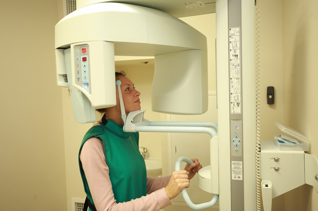
An x-ray of the maxillary sinuses is performed by a specialist in a clinical setting, takes several minutes, and it will take about another half an hour to receive the results of the analysis. The procedure itself is very simple and painless for the patient, it does not require any specific preparations and restrictions.
The specialist can perform x-rays of the sinuses in different positions of the patient, including standing. Most often, the patient is asked to lie face down on the table and open his mouth wide. This is done in order to get the clearest possible image of the sinuses without unnecessary shadows. A few minutes - and the procedure is completed. No further action is required from the patient. The finished radiographic image with the results of the decoding is transferred to the attending physician for selection and appointment of the appropriate one.
Everyone can perform x-rays of the maxillary sinuses as prescribed by a doctor, but pregnant women should avoid this procedure.
However, according to vital indications and with the permission of a doctor, an x-ray can be done.Re-appointment for x-ray examination of the sinuses is possible after a comprehensive treatment of the disease to control its quality and verify a complete cure for the problem.
Most often, a second x-ray is prescribed after the walls of the maxillary sinuses are punctured and washed out of the contents. After taking all the necessary physiotherapeutic procedures and medications, the doctor must verify the effectiveness of the measures taken and the complete destruction of the inflammatory process.
 Normally, an x-ray of the maxillary sinuses shows a clear outline of the sinuses without various pathological changes. In the picture, the patient's skull bones and teeth look light, white, and the soft tissues seem to disappear.
The cavities and sinuses look like dark spots. As a comparative characteristic of the permeability and depth of the sinus, the degree of darkening of the orbits is used. Under normal conditions, the intensity of staining of the maxillary sinuses and eye sockets should be approximately the same. The walls of the frontal and maxillary sinuses should have even outlines, there should not be any shadows around them. The appearance of darkening inside the sinus contour of different widths and degrees of intensity demonstrates the presence of inflamed mucous membranes.
The thickness of the opacity is the size of the enlarged mucosa and can be used to determine the degree of development of the inflammatory process.
In the absence of accumulation of fluid in the sinuses, such blackouts indicate that the inflammatory process has just begun, and sinusitis has not yet developed. This is the most favorable time to start high-quality complex treatment of the disease. Normally, there should be no changes in the thickness of the walls of the mucous membranes. Such a person is absolutely healthy.
An important property is its ability to show the structure of the bones of the skull. Sometimes a person has been looking for the cause of his chronic runny nose for years, and only after receiving an x-ray image does it become clear. Most often, this is a curvature of the nasal septum, the presence of or adenoids, as well as various others. Timely performance of an x-ray examination can detect in the early stages and prevent the development of many different, up to very serious, life-threatening patients. Therefore, you should not refuse the doctor's recommendation to take a picture of the facial part of the skull - it will not be able to harm you, but will only bring benefits.
Possible diseases

On an x-ray, the doctor can see inflammation, diseases and neoplasms in the maxillary sinuses
When performing x-rays of the maxillary and frontal sinuses, the following diseases and problems can be detected:
- Deviation of the nasal septum.
- Defects in the structure of the bones of the skull.
- Traces of injuries and fractures, displacement of individual sections of the facial skeleton.
- The presence of foreign bodies in the nose, nasopharynx and sinuses, which very often happens with young children.
- Inflammation of the mucous membranes of the nasal sinuses (on a radiographic image it is reflected in the form of a narrow strip of darkening along the outlines of the nasal sinuses).
- The presence of neoplasms of various types (cysts, polyps, adenoids, other types of benign and malignant tumors).
Diseases caused by acute or chronic inflammatory processes. In the picture, they are reflected in the form of blackouts along the outlines of the sinuses (inflammation of the mucous membranes) and light spots at the bottom of the sinuses, showing the level of accumulation of fluid or pus. If the sinuses turn out to be a bright spot in the picture, this indicates that they are almost “clogged” to the top with mucopurulent contents. Such a diagnosis indicates a strong and most likely long-standing inflammatory process that requires rapid action to treat the patient.
An experienced specialist will immediately see the presence of a variety of problems in just one x-ray image.
For example, the presence of fluid in the sinuses can be compared by a radiologist with the volume of fluid poured into a cup or glass. In the same way, it accumulates in the nasal and frontal sinuses in the presence of an inflammatory process provoked by a severe runny nose. Each of the fluids has its own density, so a good specialist in the level of white in the picture can tell what is contained in the patient's sinuses. If the white color appears as a light whitish haze, this may mean that the liquid is quite transparent, mucous. And this, in turn, testifies to the recent beginning of the process.
More information about sinusitis can be found in the video:
If the picture shows areas of a fairly dense white color, the doctor will logically assume that the inflammatory process has already gone far and very dense thick mucus has accumulated in the maxillary and frontal sinuses of the patient, possibly having a purulent character. Such data indicate the presence of advanced sinusitis, which can seriously threaten the patient's health.
Thus, only one radiographic image can give the doctor a lot of useful information about the condition of the sinuses of the patient's skull.